Copyright
©The Author(s) 2018.
World J Gastroenterol. Sep 21, 2018; 24(35): 3980-3999
Published online Sep 21, 2018. doi: 10.3748/wjg.v24.i35.3980
Published online Sep 21, 2018. doi: 10.3748/wjg.v24.i35.3980
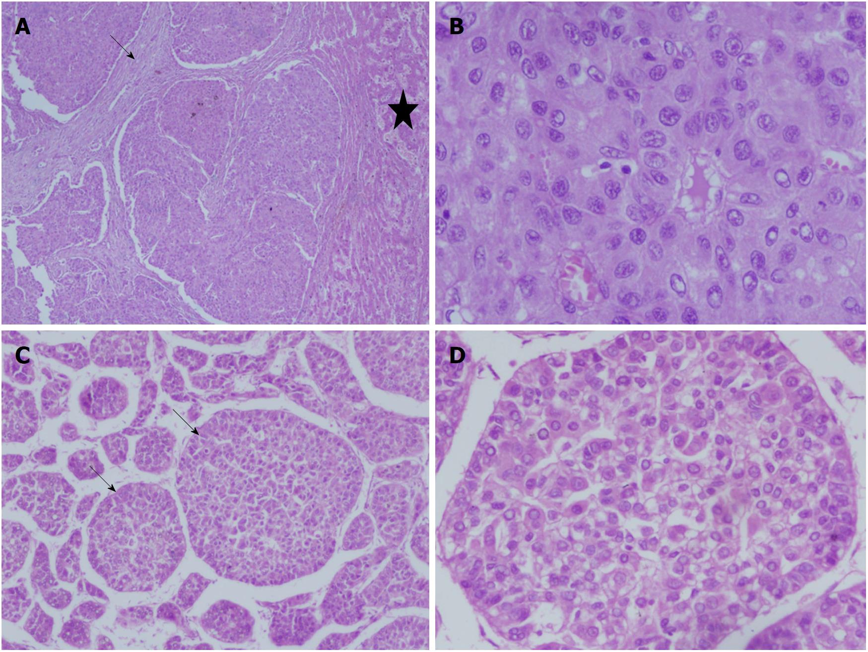
Figure 4 Liver histology images in children with hepatocellular carcinoma.
A and B: 15 yr old boy with HBeAg negative chronic hepatitis-B, DNA 4 log, precore mutant, AFP = 150000 ng/mL, with a large HCC in right lobe, with portal vein thrombosis, BCLC stage C, Child Pugh A, underwent right hepatectomy. Low power view showing tumor nodules separated by fibrous bands (arrow), adjacent non-malignant parenchyma (star) (100 ×, HE stain) (A). High power view showing malignant nuclear details with prominent nucleoli and eoisnophilic cytoplasm (400 ×, HE stain) (B); C and D: 14 and half years old boy with chronic hepatitis-B (Immunoclearance phase), HBeAg+, DNA 5 log, AFP = 3 ng/mL (normal), with a segment 3 lesion (2.5 cm × 2.4 cm), BCLC stage A, underwent non-anatomic resection. Low power view of tumor showing broadened trabeculae (arrows) (100 ×, HE stain) (C). Trabeculae with malignant cytological features - nuclear atypia and anisocytosis (200 ×, HE stain) (D).
- Citation: Khanna R, Verma SK. Pediatric hepatocellular carcinoma. World J Gastroenterol 2018; 24(35): 3980-3999
- URL: https://www.wjgnet.com/1007-9327/full/v24/i35/3980.htm
- DOI: https://dx.doi.org/10.3748/wjg.v24.i35.3980


