Copyright
©The Author(s) 2018.
World J Gastroenterol. Jan 21, 2018; 24(3): 408-414
Published online Jan 21, 2018. doi: 10.3748/wjg.v24.i3.408
Published online Jan 21, 2018. doi: 10.3748/wjg.v24.i3.408
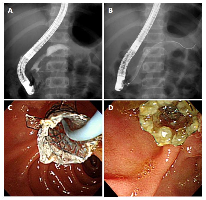
Figure 2 Endoscopic retrograde pancreatography images for case 3.
A: Pancreatography image showing a pancreatic duct stricture in the pancreatic neck before the insertion of the fully covered self-expandable metal stent (FCSEMS). B and C: An FCSEMS with flaps placed through the narrow pancreatic duct. D: The stent lumen showing patency at 6 mo after FCSEMS placement.
- Citation: Jeong IS, Lee SH, Oh SH, Park DH, Kim KM. Metal stents placement for refractory pancreatic duct stricture in children. World J Gastroenterol 2018; 24(3): 408-414
- URL: https://www.wjgnet.com/1007-9327/full/v24/i3/408.htm
- DOI: https://dx.doi.org/10.3748/wjg.v24.i3.408


