Copyright
©The Author(s) 2018.
World J Gastroenterol. Aug 7, 2018; 24(29): 3239-3249
Published online Aug 7, 2018. doi: 10.3748/wjg.v24.i29.3239
Published online Aug 7, 2018. doi: 10.3748/wjg.v24.i29.3239
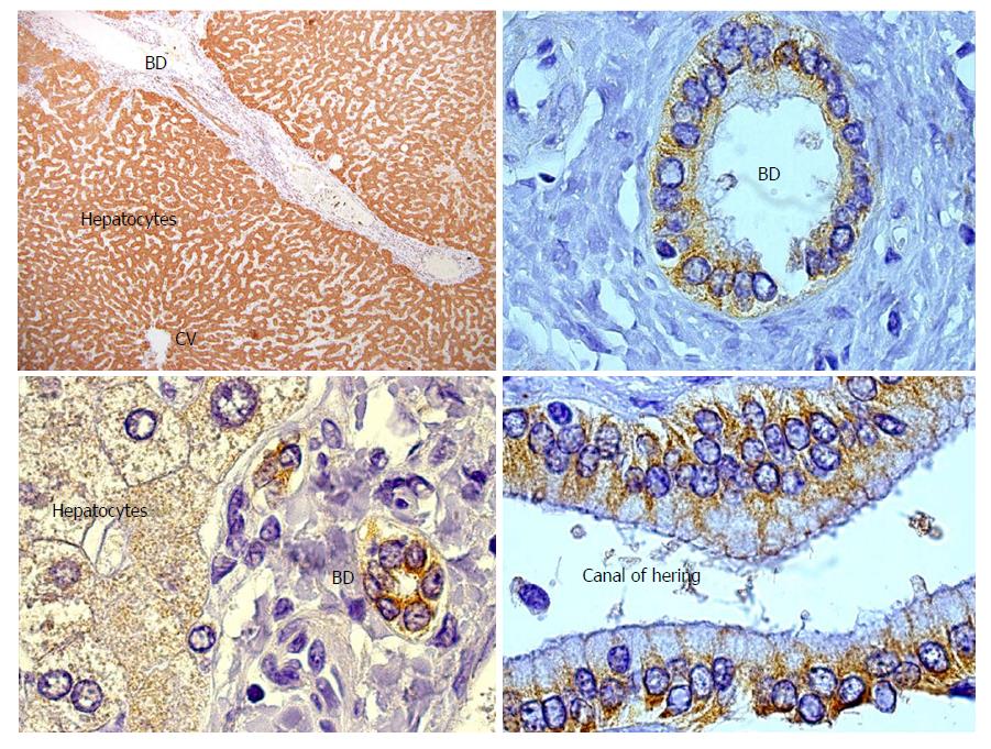
Figure 1 GSTT1 expression in human liver.
On the left part of the figure the hepatic parenchyma shows homogeneous staining of the cytoplasm of hepatocytes as well as epithelial cells of the bile ducts in the portal area. On the right part, a detail of the cytoplasmic staining of cholangiocytes and the Canal of Hering cells, considered a niche of hepatic progenitor cells. CV: Central vein; BD: Bile duct.
- Citation: Aguilera I, Aguado-Dominguez E, Sousa JM, Nuñez-Roldan A. Rethinking de novo immune hepatitis, an old concept for liver allograft rejection: Relevance of glutathione S-transferase T1 mismatch. World J Gastroenterol 2018; 24(29): 3239-3249
- URL: https://www.wjgnet.com/1007-9327/full/v24/i29/3239.htm
- DOI: https://dx.doi.org/10.3748/wjg.v24.i29.3239


