Copyright
©The Author(s) 2018.
World J Gastroenterol. Jul 14, 2018; 24(26): 2853-2866
Published online Jul 14, 2018. doi: 10.3748/wjg.v24.i26.2853
Published online Jul 14, 2018. doi: 10.3748/wjg.v24.i26.2853
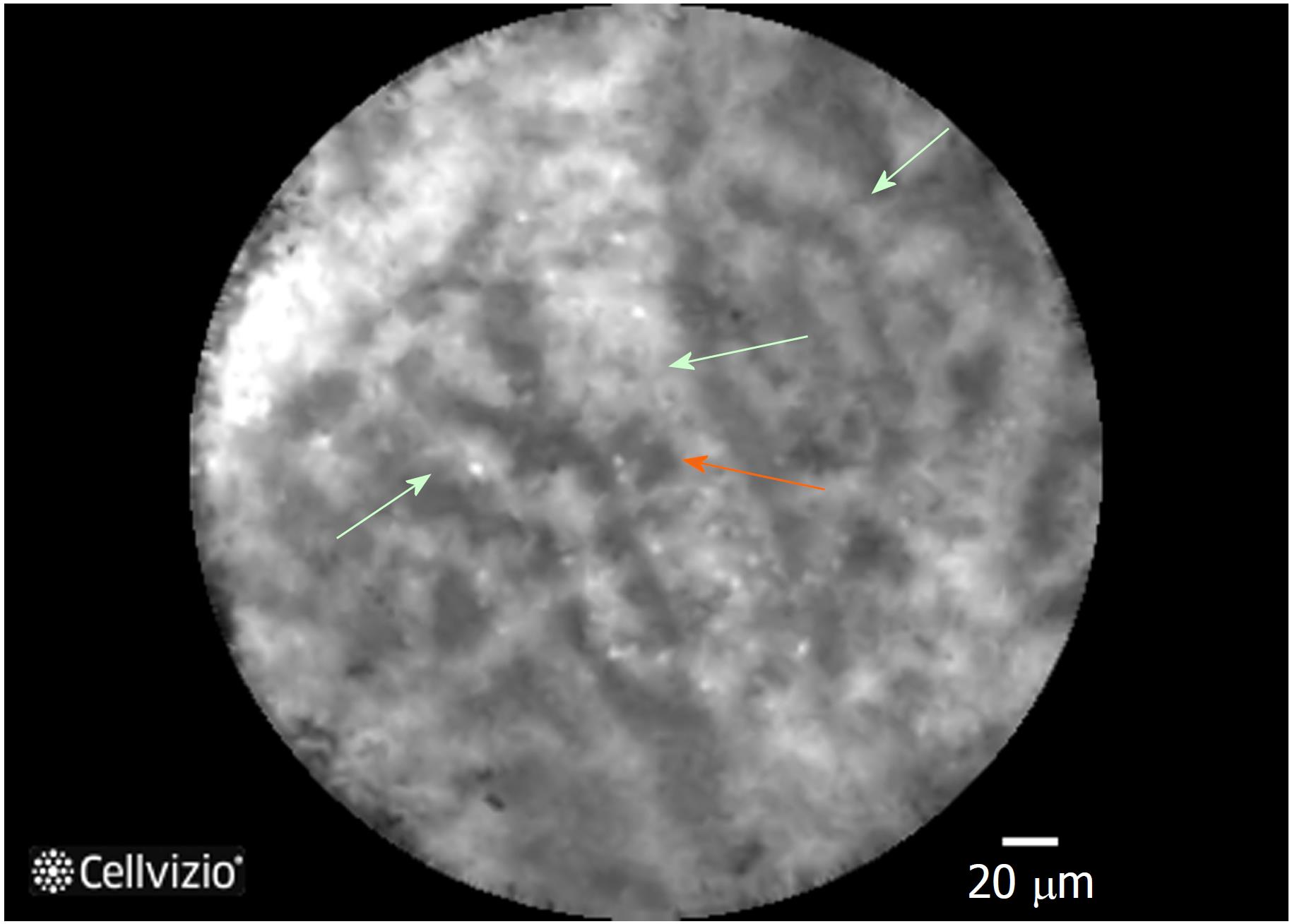
Figure 4 Needle-based confocal laser endomicroscopy image of the superficial vascular network pattern of a serous cystadenoma.
Multiple interconnected vessels (green arrows). Red cells inside displayed as black structures (orange arrow).
- Citation: Alvarez-Sánchez MV, Napoléon B. New horizons in the endoscopic ultrasonography-based diagnosis of pancreatic cystic lesions. World J Gastroenterol 2018; 24(26): 2853-2866
- URL: https://www.wjgnet.com/1007-9327/full/v24/i26/2853.htm
- DOI: https://dx.doi.org/10.3748/wjg.v24.i26.2853


