Copyright
©The Author(s) 2018.
World J Gastroenterol. Jun 14, 2018; 24(22): 2348-2362
Published online Jun 14, 2018. doi: 10.3748/wjg.v24.i22.2348
Published online Jun 14, 2018. doi: 10.3748/wjg.v24.i22.2348
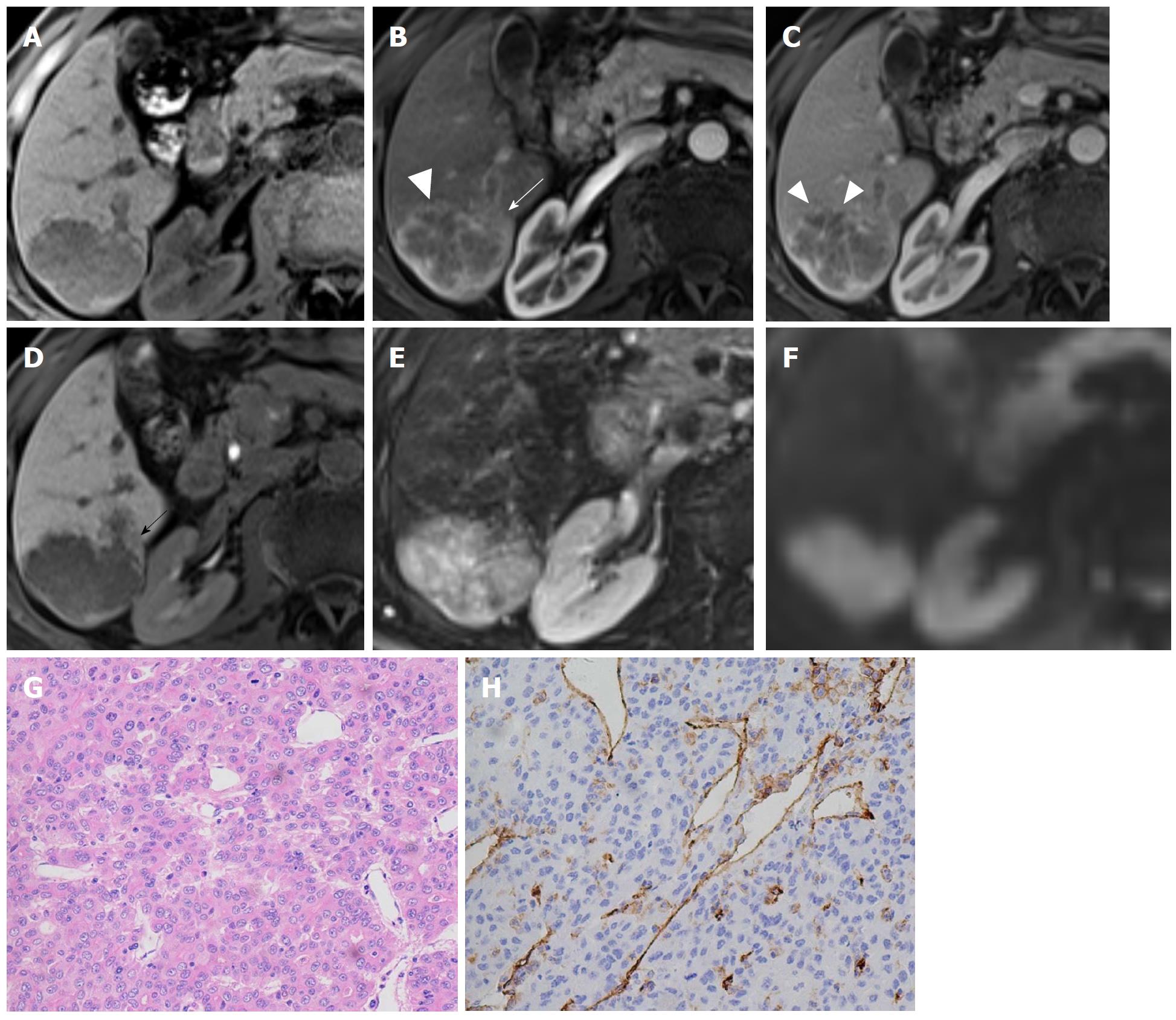
Figure 3 Hepatocellular carcinoma in a 71-year-old male with recognized cirrhosis.
Gd-EOB-DTPA-enhanced MR image demonstrates a 5.3 cm lobulated HCC in right posterior section of liver. The lesion shows peritumor enhancement in arterial phase (B, white arrow) and peritumor hypointense (D, black arrow) in hepatobiliary phase. Capsular disruption and non-smooth tumor margin are present (white triangles) in arterial phase (B) and portal venous phase (C). The lesion was histopathologically proven to be Edmonson-Steiner III grade with hematoxylin-eosin (HE) staining at 200 × magnification (G). Prominent microvascular invasion was detected at 200 × magnification with CD31 immunohistochemical staining (H).
- Citation: Jiang HY, Chen J, Xia CC, Cao LK, Duan T, Song B. Noninvasive imaging of hepatocellular carcinoma: From diagnosis to prognosis. World J Gastroenterol 2018; 24(22): 2348-2362
- URL: https://www.wjgnet.com/1007-9327/full/v24/i22/2348.htm
- DOI: https://dx.doi.org/10.3748/wjg.v24.i22.2348


