Copyright
©The Author(s) 2018.
World J Gastroenterol. Jan 14, 2018; 24(2): 237-247
Published online Jan 14, 2018. doi: 10.3748/wjg.v24.i2.237
Published online Jan 14, 2018. doi: 10.3748/wjg.v24.i2.237
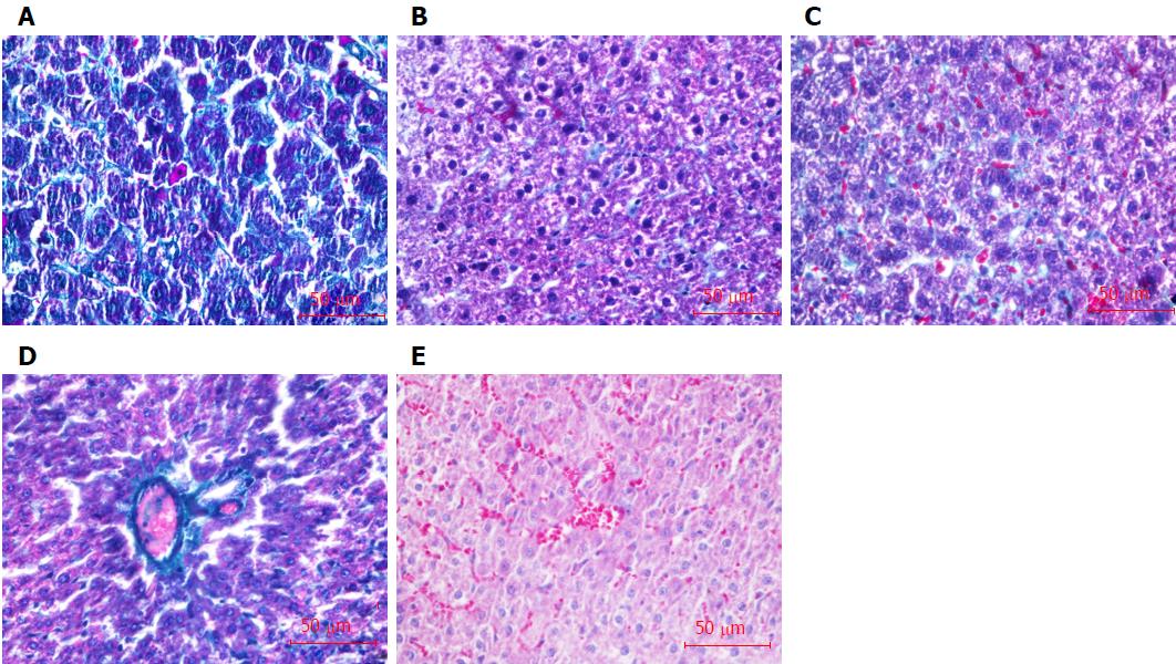
Figure 5 Collagen formation in the liver shown by masson staining (green).
A: Normal group; B: Model group; C: Bone marrow-derived hepatocyte stem cells group; D: Bone marrow-derived endothelial progenitor cells group (BM-EPCs); E: BM-EPCs/BDHSCs group. Bars: 50 μm.
- Citation: Lan L, Liu R, Qin LY, Cheng P, Liu BW, Zhang BY, Ding SZ, Li XL. Transplantation of bone marrow-derived endothelial progenitor cells and hepatocyte stem cells from liver fibrosis rats ameliorates liver fibrosis. World J Gastroenterol 2018; 24(2): 237-247
- URL: https://www.wjgnet.com/1007-9327/full/v24/i2/237.htm
- DOI: https://dx.doi.org/10.3748/wjg.v24.i2.237


