Copyright
©The Author(s) 2018.
World J Gastroenterol. May 14, 2018; 24(18): 2009-2023
Published online May 14, 2018. doi: 10.3748/wjg.v24.i18.2009
Published online May 14, 2018. doi: 10.3748/wjg.v24.i18.2009
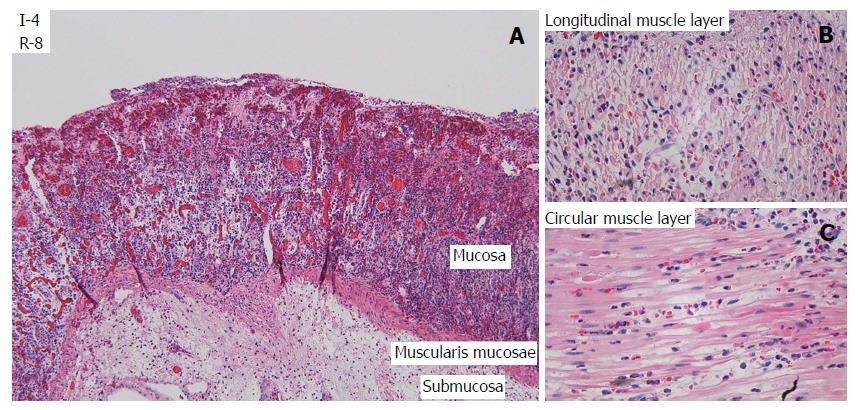
Figure 3 Light microscopy of selected structures of the jejunum after 4 h of ischemia and 8 h of reperfusion.
A: Mucosa and submucosa (HE, × 10), showing necrotic villi, total loss of crypt epithelium, shrinkage of myocytes in the muscularis mucosae, and edema in the submucosa. B: Longitudinal (outer) layer of the muscularis propria, showing edema and extensive shrinkage and loss of myocytes (HE, × 60). C: Circular (inner) layer of the muscularis propria, showing edema and extensive myocyte damage (HE, × 60).
- Citation: Strand-Amundsen RJ, Reims HM, Reinholt FP, Ruud TE, Yang R, Høgetveit JO, Tønnessen TI. Ischemia/reperfusion injury in porcine intestine - Viability assessment. World J Gastroenterol 2018; 24(18): 2009-2023
- URL: https://www.wjgnet.com/1007-9327/full/v24/i18/2009.htm
- DOI: https://dx.doi.org/10.3748/wjg.v24.i18.2009


