Copyright
©The Author(s) 2018.
World J Gastroenterol. Apr 7, 2018; 24(13): 1419-1428
Published online Apr 7, 2018. doi: 10.3748/wjg.v24.i13.1419
Published online Apr 7, 2018. doi: 10.3748/wjg.v24.i13.1419
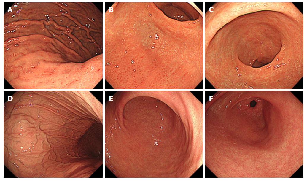
Figure 3 Representative endoscopic findings of negative-high titer antibody cases.
A case with Helicobacter pylori infection; 81-year-old woman with antibody titer of 4.7 U/mL, UBT of 7.3 per mil, and Kyoto classification score of 5 (A-C). A: Greater curvature of the body of the stomach. Enlarged folds and redness are present; B: Lower body of the stomach. Endoscopic atrophic border lies in the anterior wall and greater curvature. Redness is present in the greater curvature; C: Antrum. Intestinal metaplasia is present in the lesser curvature. The mucosa is atrophic. A case without H. pylori infection; 31-year-old man with antibody titer of 5.7 U/mL, UBT of 1.2 per mil, and Kyoto classification score of 0 (D-F); D: The greater curvature of the body of the stomach. Regular arrangement of collecting venules and fundic gland polyps are present; E: Lower body of the stomach. Atrophy and redness are absent; F: Antrum. Intestinal metaplasia and atrophy are absent. UBT: Urea breath test.
- Citation: Toyoshima O, Nishizawa T, Arita M, Kataoka Y, Sakitani K, Yoshida S, Yamashita H, Hata K, Watanabe H, Suzuki H. Helicobacter pylori infection in subjects negative for high titer serum antibody. World J Gastroenterol 2018; 24(13): 1419-1428
- URL: https://www.wjgnet.com/1007-9327/full/v24/i13/1419.htm
- DOI: https://dx.doi.org/10.3748/wjg.v24.i13.1419


