Copyright
©The Author(s) 2018.
World J Gastroenterol. Apr 7, 2018; 24(13): 1419-1428
Published online Apr 7, 2018. doi: 10.3748/wjg.v24.i13.1419
Published online Apr 7, 2018. doi: 10.3748/wjg.v24.i13.1419
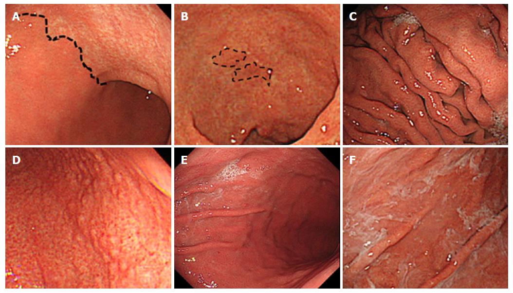
Figure 1 Endoscopic findings related to Helicobacter pylori infection.
A: Atrophy is diagnosed based on the vascular pattern and rugal atrophy. The dotted line indicates an atrophic border in the anterior wall of the body (43-year-old woman; antibody titer: 4.3 U/mL; UBT: 55.3 per mil; Kyoto classification score: 2); B: Intestinal metaplasia is visible as grayish-whitish, slightly opalescent patches. The dotted line indicates the extent of the lesions in the lesser curvature of the antrum (81-year-old woman; antibody titer: 4.7 U/mL; UBT: 7.3 per mil; Kyoto classification score: 5); C: An enlarged fold is defined as that which is 5 mm or more in diameter. Enlarged folds are present in the greater curvature of the body (56-year-old man; antibody titer: 3.8 U/mL; UBT: 7.0 per mil; Kyoto classification score: 3); D: Nodularity is characterized by the appearance of multiple whitish elevated lesions mainly in the pyloric gland mucosa. Nodularity is present in the antrum (28-year-old man; antibody titer: 9.4 U/mL; UBT: 3.6 per mil; Kyoto classification score: 2); E: Redness refers to uniform redness involving the entire fundic gland mucosa. Redness is visible in the greater curvature of the body (44-year-old man; antibody titer: 8.7 U/mL; UBT: 26.5 per mil; Kyoto classification score: 3); F: Sticky mucus refers to grayish or yellowish mucus adhering to the mucosal surface. There is sticky mucus in the greater curvature of the body (70-year-old woman; antibody titer: 6.5 U/mL; UBT: 26.4 per mil; Kyoto classification score: 4). UBT: Urea breath test.
- Citation: Toyoshima O, Nishizawa T, Arita M, Kataoka Y, Sakitani K, Yoshida S, Yamashita H, Hata K, Watanabe H, Suzuki H. Helicobacter pylori infection in subjects negative for high titer serum antibody. World J Gastroenterol 2018; 24(13): 1419-1428
- URL: https://www.wjgnet.com/1007-9327/full/v24/i13/1419.htm
- DOI: https://dx.doi.org/10.3748/wjg.v24.i13.1419


