Copyright
©The Author(s) 2017.
World J Gastroenterol. Feb 28, 2017; 23(8): 1353-1366
Published online Feb 28, 2017. doi: 10.3748/wjg.v23.i8.1353
Published online Feb 28, 2017. doi: 10.3748/wjg.v23.i8.1353
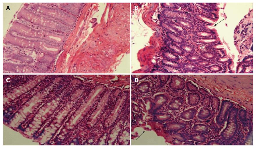
Figure 4 Representative histological appearance of rat colonic mucosa in the non-colitic group (A), colitic group (B), Cd-EtOHE (C), and Cd-HexP (D).
Aumento magnification × 40 (*goblet cells; → ulceration region; 50 μm). Colonic tissue sections were stained with hematoxylin and eosin, and observed under light microscope (magnification × 40). Cd: Combretum duarteanum; HexP: Hexane phase; EtOHE: Ethanolic extract.
- Citation: de Morais Lima GR, Machado FDF, Périco LL, de Faria FM, Luiz-Ferreira A, Souza Brito ARM, Pellizzon CH, Hiruma-Lima CA, Tavares JF, Barbosa Filho JM, Batista LM. Anti-inflammatory intestinal activity of Combretum duarteanum Cambess. in trinitrobenzene sulfonic acid colitis model. World J Gastroenterol 2017; 23(8): 1353-1366
- URL: https://www.wjgnet.com/1007-9327/full/v23/i8/1353.htm
- DOI: https://dx.doi.org/10.3748/wjg.v23.i8.1353


