Copyright
©The Author(s) 2017.
World J Gastroenterol. Feb 14, 2017; 23(6): 1113-1118
Published online Feb 14, 2017. doi: 10.3748/wjg.v23.i6.1113
Published online Feb 14, 2017. doi: 10.3748/wjg.v23.i6.1113
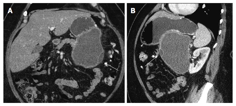
Figure 2 Coronal (A) and sagittal (B) images in portal venous phase show the same lesion abutting the stomach in its superior aspect.
Thin septations and small loculations are again noted (arrows).
- Citation: Mederos MA, Villafañe N, Dhingra S, Farinas C, McElhany A, Fisher WE, Van Buren II G. Pancreatic endometrial cyst mimics mucinous cystic neoplasm of the pancreas. World J Gastroenterol 2017; 23(6): 1113-1118
- URL: https://www.wjgnet.com/1007-9327/full/v23/i6/1113.htm
- DOI: https://dx.doi.org/10.3748/wjg.v23.i6.1113


