Copyright
©The Author(s) 2017.
World J Gastroenterol. Feb 14, 2017; 23(6): 1106-1112
Published online Feb 14, 2017. doi: 10.3748/wjg.v23.i6.1106
Published online Feb 14, 2017. doi: 10.3748/wjg.v23.i6.1106
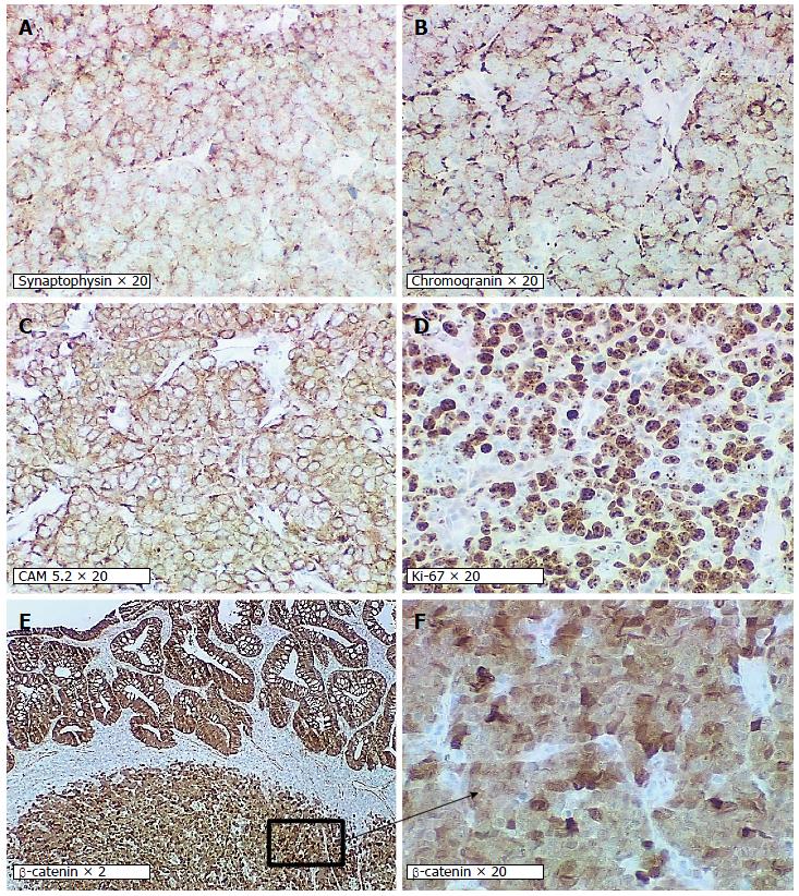
Figure 3 High-grade large-cell neuroendocrine carcinoma.
Immunohistochemical profile of the tumor showing positive staining for synaptophysin (A) and chromogranin (B) in the neuroendocrine carcinoma component. Both the glandular and neuroendocrine components stained positive for CAM5.2 (C) and β-catenin (E and F). The Ki-67 labeling index is approximately 75% positive in stained tumor cells (D).
- Citation: Soliman ML, Tiwari A, Zhao Q. Coexisting tubular adenoma with a neuroendocrine carcinoma of colon allowing early surgical intervention and implicating a shared stem cell origin. World J Gastroenterol 2017; 23(6): 1106-1112
- URL: https://www.wjgnet.com/1007-9327/full/v23/i6/1106.htm
- DOI: https://dx.doi.org/10.3748/wjg.v23.i6.1106


