Copyright
©The Author(s) 2017.
World J Gastroenterol. Feb 7, 2017; 23(5): 919-925
Published online Feb 7, 2017. doi: 10.3748/wjg.v23.i5.919
Published online Feb 7, 2017. doi: 10.3748/wjg.v23.i5.919
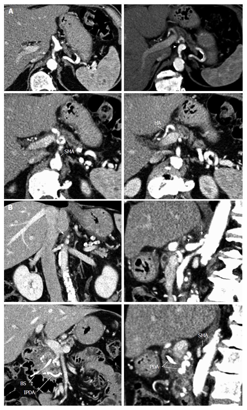
Figure 2 Features of median arcuate ligament.
Arterial phase of the axial computerized tomography (CT) scan (A), along with coronal and sagittal CT-scan reconstructions (B) showing severe stenosis of the celiac trunk from extrinsic compression by dense fibrous tissue (arrow) and poststenotic dilation of the proximal celiac trunk (arrowhead). SMA: Superior mesenteric artery; HA: Hepatic artery; PDA: Pancreaticoduodenal arcade; IPDA: Inferior pancreaticoduodenal artery; BS: Biliary stent.
- Citation: Guilbaud T, Ewald J, Turrini O, Delpero JR. Pancreaticoduodenectomy: Secondary stenting of the celiac trunk after inefficient median arcuate ligament release and reoperation as an alternative to simultaneous hepatic artery reconstruction. World J Gastroenterol 2017; 23(5): 919-925
- URL: https://www.wjgnet.com/1007-9327/full/v23/i5/919.htm
- DOI: https://dx.doi.org/10.3748/wjg.v23.i5.919


