Copyright
©The Author(s) 2017.
World J Gastroenterol. Dec 28, 2017; 23(48): 8671-8678
Published online Dec 28, 2017. doi: 10.3748/wjg.v23.i48.8671
Published online Dec 28, 2017. doi: 10.3748/wjg.v23.i48.8671
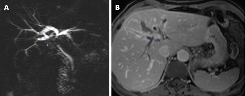
Figure 2 Preoperative magnetic resonance cholangiopancreatography from case 2 demonstrates stenosis of the proximal and middle thirds of the common bile duct, biliary confluence (arrowhead, A), and right hepatic duct and second-order biliary radicals, with retrograde biliary dilatation; a suspicious-appearing spiculated hilar lymph node is seen on transverse section (arrow, B).
- Citation: Nacif LS, Hessheimer AJ, Rodríguez Gómez S, Montironi C, Fondevila C. Infiltrative xanthogranulomatous cholecystitis mimicking aggressive gallbladder carcinoma: A diagnostic and therapeutic dilemma. World J Gastroenterol 2017; 23(48): 8671-8678
- URL: https://www.wjgnet.com/1007-9327/full/v23/i48/8671.htm
- DOI: https://dx.doi.org/10.3748/wjg.v23.i48.8671


