Copyright
©The Author(s) 2017.
World J Gastroenterol. Dec 28, 2017; 23(48): 8597-8604
Published online Dec 28, 2017. doi: 10.3748/wjg.v23.i48.8597
Published online Dec 28, 2017. doi: 10.3748/wjg.v23.i48.8597
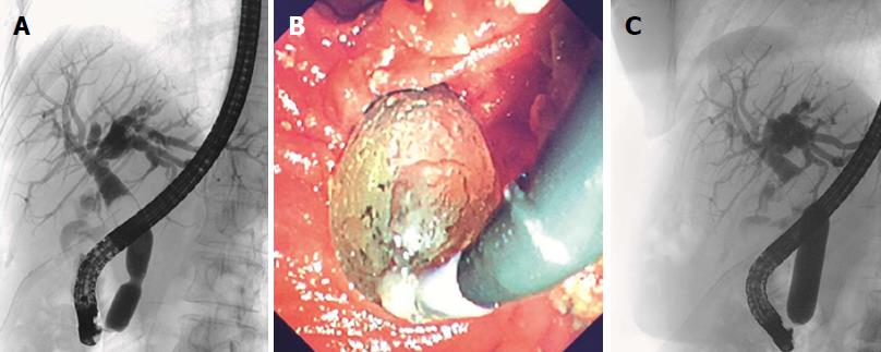
Figure 1 Demonstration of endoscopic papillary large balloon dilatation.
A: Fluoroscopic view showing balloon waist with a proximal stone; B: Endoscopic view of ampullary orifice while performing EPLBD; C: Demonstration of disappearance of balloon waist with gradual inflation of balloon.
- Citation: Aujla UI, Ladep N, Dwyer L, Hood S, Stern N, Sturgess R. Endoscopic papillary large balloon dilatation with sphincterotomy is safe and effective for biliary stone removal independent of timing and size of sphincterotomy. World J Gastroenterol 2017; 23(48): 8597-8604
- URL: https://www.wjgnet.com/1007-9327/full/v23/i48/8597.htm
- DOI: https://dx.doi.org/10.3748/wjg.v23.i48.8597


