Copyright
©The Author(s) 2017.
World J Gastroenterol. Dec 14, 2017; 23(46): 8248-8255
Published online Dec 14, 2017. doi: 10.3748/wjg.v23.i46.8248
Published online Dec 14, 2017. doi: 10.3748/wjg.v23.i46.8248
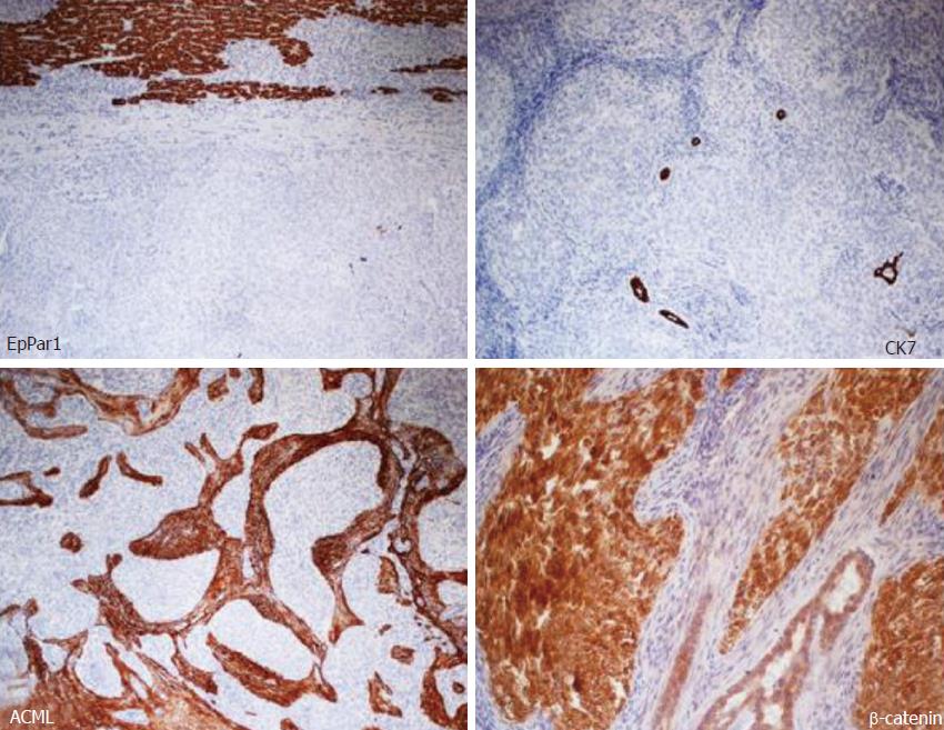
Figure 5 Immunohistochemical stains showing neoplasm negativity for hepatocyte paraffin 1 (EpPar1) (counterstained normal liver parenchyma), negativity for CK7 (highlighted entrapped bile ducts between the tumor cell), stromal positivity for ACML and positivity for β-catenin (both membrane and nuclear).
- Citation: Meletani T, Cantini L, Lanese A, Nicolini D, Cimadamore A, Agostini A, Ricci G, Antognoli S, Mandolesi A, Guido M, Alaggio R, Giuseppetti GM, Scarpelli M, Vivarelli M, Berardi R. Are liver nested stromal epithelial tumors always low aggressive? World J Gastroenterol 2017; 23(46): 8248-8255
- URL: https://www.wjgnet.com/1007-9327/full/v23/i46/8248.htm
- DOI: https://dx.doi.org/10.3748/wjg.v23.i46.8248


