Copyright
©The Author(s) 2017.
World J Gastroenterol. Dec 7, 2017; 23(45): 8090-8096
Published online Dec 7, 2017. doi: 10.3748/wjg.v23.i45.8090
Published online Dec 7, 2017. doi: 10.3748/wjg.v23.i45.8090
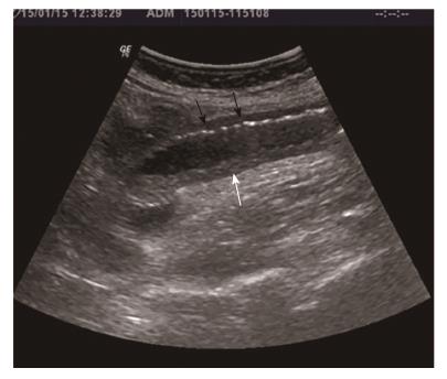
Figure 2 Sonographic features show dilated small bowel loop with absent peristalsis.
Also depicted are increased intraluminal secretions within the ischemic small bowel segment (white arrow), slight mural thickening and intramural gas (black arrows).
- Citation: Mantzoros I, Savvala NA, Ioannidis O, Parpoudi S, Loutzidou L, Kyriakidou D, Cheva A, Intzos V, Tsalis K. Midgut neuroendocrine tumor presenting with acute intestinal ischemia. World J Gastroenterol 2017; 23(45): 8090-8096
- URL: https://www.wjgnet.com/1007-9327/full/v23/i45/8090.htm
- DOI: https://dx.doi.org/10.3748/wjg.v23.i45.8090


