Copyright
©The Author(s) 2017.
World J Gastroenterol. Nov 7, 2017; 23(41): 7478-7488
Published online Nov 7, 2017. doi: 10.3748/wjg.v23.i41.7478
Published online Nov 7, 2017. doi: 10.3748/wjg.v23.i41.7478
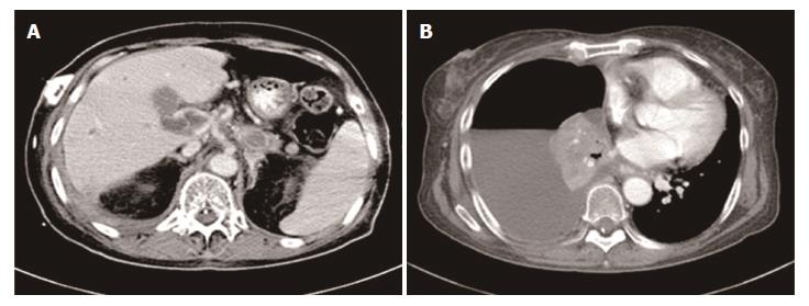
Figure 6 Disease progression.
CT scan revealed several metastatic lesions in the liver (A) and pleural effusion (B). CT: Computed tomography.
- Citation: Li CM, Liu ZC, Bao YT, Sun XD, Wang LL. Extraordinary response of metastatic pancreatic cancer to apatinib after failed chemotherapy: A case report and literature review. World J Gastroenterol 2017; 23(41): 7478-7488
- URL: https://www.wjgnet.com/1007-9327/full/v23/i41/7478.htm
- DOI: https://dx.doi.org/10.3748/wjg.v23.i41.7478


