Copyright
©The Author(s) 2017.
World J Gastroenterol. Nov 7, 2017; 23(41): 7347-7358
Published online Nov 7, 2017. doi: 10.3748/wjg.v23.i41.7347
Published online Nov 7, 2017. doi: 10.3748/wjg.v23.i41.7347
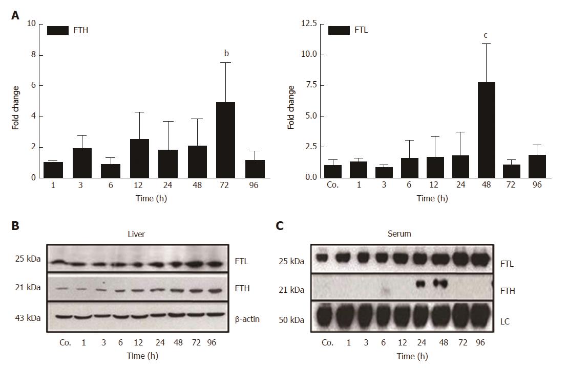
Figure 4 Changes in hepatic and serum levels of ferritin heavy chain and ferritin light chain after thioacetamide-induced liver damage.
A: Significant (bP ≤ 0.01; cP ≤ 0.001) fold-change of hepatic mRNA expression of ferritin heavy chain (FTH) and ferritin light chain (FTL) in TAA-injected animals vs controls (Co.) after 72 h and 48 h, respectively, analyzed by qPCR and normalized against the house-keeping gene UBC. Results represent mean values ± SEM of each four animals per group. Controls were set as 1; B, C: Western blot analysis of FTL and FTH in total liver and in serum of control (Co.) and TAA-injected animals at indicated time points. β-actin (about 43 kDa) was used as loading control for liver tissue, while in serum the loading control (LC) represents an internal control (about 50 kDa).
- Citation: Malik IA, Wilting J, Ramadori G, Naz N. Reabsorption of iron into acutely damaged rat liver: A role for ferritins. World J Gastroenterol 2017; 23(41): 7347-7358
- URL: https://www.wjgnet.com/1007-9327/full/v23/i41/7347.htm
- DOI: https://dx.doi.org/10.3748/wjg.v23.i41.7347


