Copyright
©The Author(s) 2017.
World J Gastroenterol. Oct 28, 2017; 23(40): 7211-7220
Published online Oct 28, 2017. doi: 10.3748/wjg.v23.i40.7211
Published online Oct 28, 2017. doi: 10.3748/wjg.v23.i40.7211
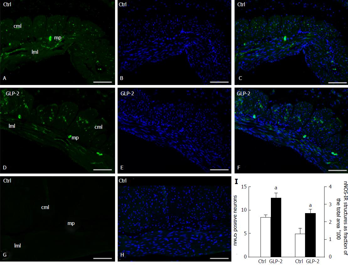
Figure 6 Neuronal nitric oxide synthase-immunoreactivity in control and glucagon-like peptide-2 treated specimens.
In control (A) and glucagon-like peptide-2 (GLP-2) treated (D) specimens, neuronal nitric oxide synthase-immunoreactivity (nNOS-IR) (green) is present in the soma of some myenteric neurons and in several nerve fibers. DAPI fluorescent staining for DNA (blue) identified the nuclei of both smooth muscle and neural cells (B-E). In C and F, merged images of nNOS and Bisbenzimide Hoechst Trihydrochloride (BHT) fluorescent staining are shown. When the primary antibody is omitted (G), only the BHT labelling is observed (H). The quantitative analysis showed a significant increase (I) in nNOS-IR myenteric neurons (left side) and in IR fibers in the entire muscle coat (right side). aP < 0.05 vs controls (unpaired Student's t-test). mp: Myenteric plexus; cml: Circular muscle layer; lml: Longitudinal muscle layer. Bar: A-H = 10 μm.
- Citation: Garella R, Idrizaj E, Traini C, Squecco R, Vannucchi MG, Baccari MC. Glucagon-like peptide-2 modulates the nitrergic neurotransmission in strips from the mouse gastric fundus. World J Gastroenterol 2017; 23(40): 7211-7220
- URL: https://www.wjgnet.com/1007-9327/full/v23/i40/7211.htm
- DOI: https://dx.doi.org/10.3748/wjg.v23.i40.7211


