Copyright
©The Author(s) 2017.
World J Gastroenterol. Oct 28, 2017; 23(40): 7211-7220
Published online Oct 28, 2017. doi: 10.3748/wjg.v23.i40.7211
Published online Oct 28, 2017. doi: 10.3748/wjg.v23.i40.7211
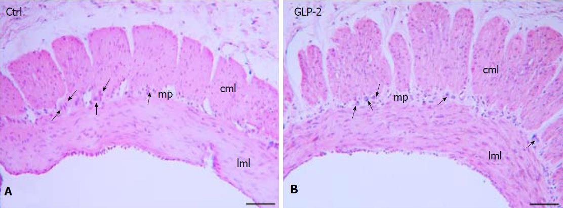
Figure 5 H&E staining of gastric fundus strip.
The longitudinal and circular muscle layers (lml, cml) and the myenteric plexus (mp) are shown both in control (A) and in GLP-2 treated (B) specimens. The arrows indicated some myenteric neurons. Bar: A, B = 10 μm.
- Citation: Garella R, Idrizaj E, Traini C, Squecco R, Vannucchi MG, Baccari MC. Glucagon-like peptide-2 modulates the nitrergic neurotransmission in strips from the mouse gastric fundus. World J Gastroenterol 2017; 23(40): 7211-7220
- URL: https://www.wjgnet.com/1007-9327/full/v23/i40/7211.htm
- DOI: https://dx.doi.org/10.3748/wjg.v23.i40.7211


