Copyright
©The Author(s) 2017.
World J Gastroenterol. Jan 28, 2017; 23(4): 735-739
Published online Jan 28, 2017. doi: 10.3748/wjg.v23.i4.735
Published online Jan 28, 2017. doi: 10.3748/wjg.v23.i4.735
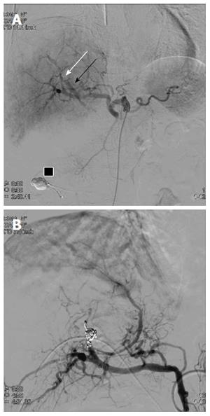
Figure 4 The 2nd digital subtraction angiography image.
A: Selective digital subtraction angiography image of the common hepatic artery showing an arterio-biliary fistula (white arrow) between A8 and the common bile duct (black arrow). Embolic material that migrated into the afferent loop is present (black box); B: Proximal A8 embolization obliterated the pseudoaneurysm. Leakage of contrast medium was completely controlled after embolization.
- Citation: Yasuda M, Sato H, Koyama Y, Sakakida T, Kawakami T, Nishimura T, Fujii H, Nakatsugawa Y, Yamada S, Tomatsuri N, Okuyama Y, Kimura H, Ito T, Morishita H, Yoshida N. Late-onset severe biliary bleeding after endoscopic pigtail plastic stent insertion. World J Gastroenterol 2017; 23(4): 735-739
- URL: https://www.wjgnet.com/1007-9327/full/v23/i4/735.htm
- DOI: https://dx.doi.org/10.3748/wjg.v23.i4.735


