Copyright
©The Author(s) 2017.
World J Gastroenterol. Oct 14, 2017; 23(38): 7054-7058
Published online Oct 14, 2017. doi: 10.3748/wjg.v23.i38.7054
Published online Oct 14, 2017. doi: 10.3748/wjg.v23.i38.7054
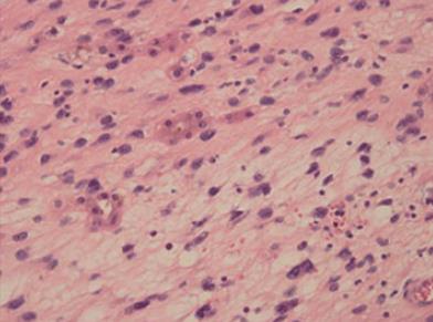
Figure 2 Histological image showing that the tumor cells were spindle shaped and located in a loose myxoid background (hematoxylin-eosin staining; original magnification, × 400).
- Citation: Wen J, Zhao W, Li C, Shen JY, Wen TF. High-grade myofibroblastic sarcoma in the liver: A case report. World J Gastroenterol 2017; 23(38): 7054-7058
- URL: https://www.wjgnet.com/1007-9327/full/v23/i38/7054.htm
- DOI: https://dx.doi.org/10.3748/wjg.v23.i38.7054


