Copyright
©The Author(s) 2017.
World J Gastroenterol. Oct 14, 2017; 23(38): 7047-7053
Published online Oct 14, 2017. doi: 10.3748/wjg.v23.i38.7047
Published online Oct 14, 2017. doi: 10.3748/wjg.v23.i38.7047
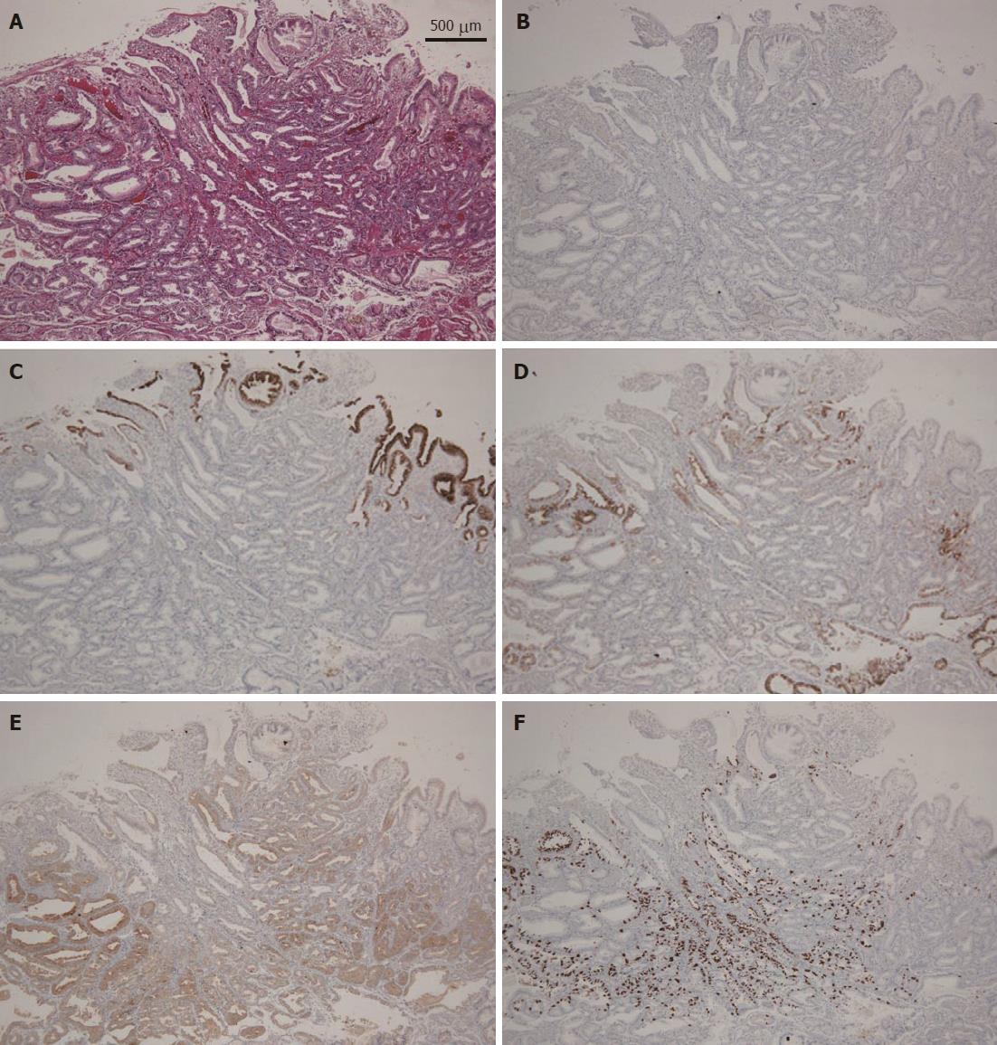
Figure 5 Histopathological and immunohistochemical findings of the endoscopic submucosal dissection-resected specimen.
A: H&E staining. There were atypical cells with mildly enlarged nuclei in the deep layer of the lamina propria mucosae. They mimicked fundic gland cells, mainly chief cells and partially parietal cells. The mucosal surface was covered completely with non-neoplastic foveolar epithelium; thus, the tumor was not exposed to the mucosal surface; B: MUC2 staining: almost negative; C: MUC5AC staining: almost negative except for normal superficial foveolar epithelium; D: MUC6 staining: partially positive and indicating a gastric phenotype; E: Pepsinogen staining: diffusely positive in the deep layer of the lamina propria mucosae, corresponding to gastric adenocarcinoma of fundic gland type, and indicating a differentiation toward chief cells; F: H/K-ATPase staining: scattered positive in the deep layer of the lamina propria mucosae, indicating a differentiation toward parietal cells focally.
- Citation: Manabe S, Mukaisho KI, Yasuoka T, Usui F, Matsuyama T, Hirata I, Boku Y, Takahashi S. Gastric adenocarcinoma of fundic gland type spreading to heterotopic gastric glands. World J Gastroenterol 2017; 23(38): 7047-7053
- URL: https://www.wjgnet.com/1007-9327/full/v23/i38/7047.htm
- DOI: https://dx.doi.org/10.3748/wjg.v23.i38.7047


