Copyright
©The Author(s) 2017.
World J Gastroenterol. Oct 14, 2017; 23(38): 7047-7053
Published online Oct 14, 2017. doi: 10.3748/wjg.v23.i38.7047
Published online Oct 14, 2017. doi: 10.3748/wjg.v23.i38.7047
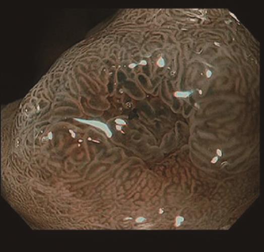
Figure 2 Narrow-band imaging with magnifying endoscopy findings.
There were few irregularities in the microvessel architecture and microsurface structure. Therefore, we diagnosed a regular microvascular pattern and a regular microsurface pattern. The depressed area at the center was expected to be a biopsy scar.
- Citation: Manabe S, Mukaisho KI, Yasuoka T, Usui F, Matsuyama T, Hirata I, Boku Y, Takahashi S. Gastric adenocarcinoma of fundic gland type spreading to heterotopic gastric glands. World J Gastroenterol 2017; 23(38): 7047-7053
- URL: https://www.wjgnet.com/1007-9327/full/v23/i38/7047.htm
- DOI: https://dx.doi.org/10.3748/wjg.v23.i38.7047


