Copyright
©The Author(s) 2017.
World J Gastroenterol. Oct 7, 2017; 23(37): 6911-6919
Published online Oct 7, 2017. doi: 10.3748/wjg.v23.i37.6911
Published online Oct 7, 2017. doi: 10.3748/wjg.v23.i37.6911
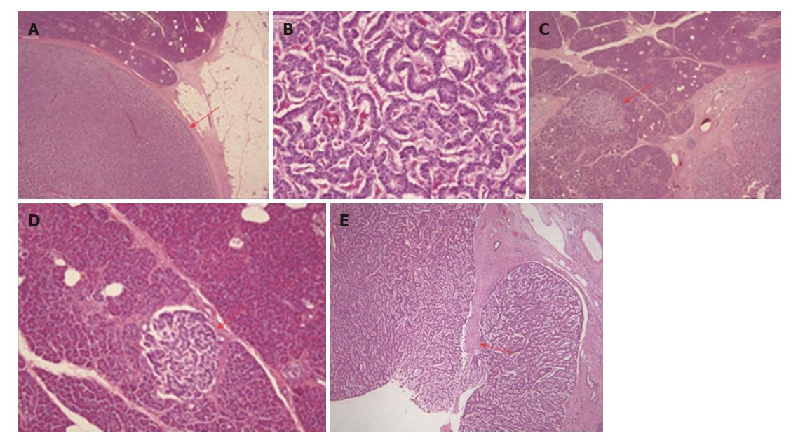
Figure 6 Microscopic findings for the resected pancreas.
A: The main lesion (red arrow) comprises cells with round nuclei arranged in sheets or rosettes (hematoxylin and eosin, × 4); B: Less than 2 mitoses per 50 high-power fields are evident (hematoxylin and eosin, × 40); C and D: The other microtumors (red arrow) show similar findings (hematoxylin and eosin; C, × 4; D, × 10); E: The peripheral rim appears broken at one site, and fibrosis (red arrow) is evident with inflammation (hematoxylin and eosin, × 10). A trace of a cystic lumen is apparent below the red arrow.
- Citation: Sagami R, Nishikiori H, Ikuyama S, Murakami K. Rupture of small cystic pancreatic neuroendocrine tumor with many microtumors. World J Gastroenterol 2017; 23(37): 6911-6919
- URL: https://www.wjgnet.com/1007-9327/full/v23/i37/6911.htm
- DOI: https://dx.doi.org/10.3748/wjg.v23.i37.6911


