Copyright
©The Author(s) 2017.
World J Gastroenterol. Oct 7, 2017; 23(37): 6911-6919
Published online Oct 7, 2017. doi: 10.3748/wjg.v23.i37.6911
Published online Oct 7, 2017. doi: 10.3748/wjg.v23.i37.6911
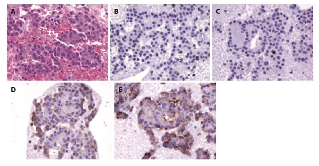
Figure 4 Histopathological findings for the specimen obtained by endoscopic ultrasound-fine needle aspiration.
A: The specimen comprises cells with round nuclei arranged in sheets or rosettes (hematoxylin and eosin, ×100); B and C: Tumor cells show negative results for CD10 (B) and CD56 (C) immunohistochemical staining (×100); D and E: Tumor cells show positive results for chromogranin A (D) and synaptophysin (E) on immunohistochemical staining (×100).
- Citation: Sagami R, Nishikiori H, Ikuyama S, Murakami K. Rupture of small cystic pancreatic neuroendocrine tumor with many microtumors. World J Gastroenterol 2017; 23(37): 6911-6919
- URL: https://www.wjgnet.com/1007-9327/full/v23/i37/6911.htm
- DOI: https://dx.doi.org/10.3748/wjg.v23.i37.6911


