Copyright
©The Author(s) 2017.
World J Gastroenterol. Oct 7, 2017; 23(37): 6911-6919
Published online Oct 7, 2017. doi: 10.3748/wjg.v23.i37.6911
Published online Oct 7, 2017. doi: 10.3748/wjg.v23.i37.6911
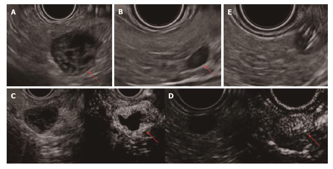
Figure 3 Findings from EUS and CH-EUS.
A: The lesion in the pancreatic tail shows a hypoechoic peripheral rim and nodule. Inside the cyst, no echoic fluid or solid structure is detected; B: A new, 7-mm microtumor in the pancreatic body, apparent only on EUS; C: The rim of the cystic lesion and nodule appear enhanced immediately after injection of contrast medium on CH-EUS; D: The 7-mm microtumor is similarly enhanced on CH-EUS; E: Endoscopic ultrasound-fine needle aspiration to biopsy the 7-mm microtumor. CH-EUS: Contrast-enhanced harmonic-endoscopic ultrasound; EUS: Endoscopic ultrasound.
- Citation: Sagami R, Nishikiori H, Ikuyama S, Murakami K. Rupture of small cystic pancreatic neuroendocrine tumor with many microtumors. World J Gastroenterol 2017; 23(37): 6911-6919
- URL: https://www.wjgnet.com/1007-9327/full/v23/i37/6911.htm
- DOI: https://dx.doi.org/10.3748/wjg.v23.i37.6911


