
















©The Author(s) 2017.
World J Gastroenterol. Oct 7, 2017; 23(37): 6902-6906
Published online Oct 7, 2017. doi: 10.3748/wjg.v23.i37.6902
Published online Oct 7, 2017. doi: 10.3748/wjg.v23.i37.6902
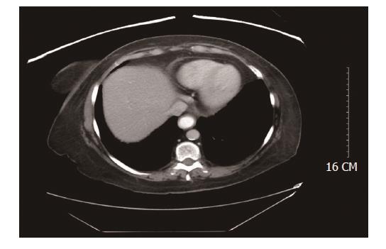
Figure 1 Computed tomography scan of the abdomen and pelvis with contrast showing enteric contrast within the dilated distal esophagus, and was suspicious for mild stricture at the gastroesophageal junction.
- Citation: Abu Ghanimeh M, Qasrawi A, Abughanimeh O, Albadarin S, Clarkston W. Achalasia after bariatric Roux-en-Y gastric bypass surgery reversal. World J Gastroenterol 2017; 23(37): 6902-6906
- URL: https://www.wjgnet.com/1007-9327/full/v23/i37/6902.htm
- DOI: https://dx.doi.org/10.3748/wjg.v23.i37.6902