Copyright
©The Author(s) 2017.
World J Gastroenterol. Oct 7, 2017; 23(37): 6833-6844
Published online Oct 7, 2017. doi: 10.3748/wjg.v23.i37.6833
Published online Oct 7, 2017. doi: 10.3748/wjg.v23.i37.6833
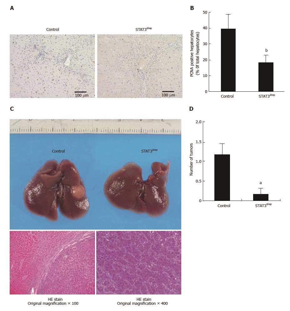
Figure 2 Hepatic STAT3 deficiency reduced hepatocyte proliferation and inhibited the development of hepatocellular carcinoma.
A: Representative immunohistochemical staining for PCNA in livers of control and STAT3Δhep mice with TAA treatment for 16 wk. PCNA positive hepatocytes were decreased in STAT3Δhep mice. Original magnification × 200. B: The number of PCNA positive hepatocytes. The ratio of PCNA positive hepatocytes to all hepatocytes was quantified in three different samples per a group. Values represent means ± standard error of the mean. bP < 0.01 (P = 0.000232) vs control. C: Gross liver appearance of control and STAT3Δhep mice with TAA treatment for 30 wk. Tumor formation with TAA treatment for 30 wk (upper panel). HE staining of representative sections of hepatocellular carcinoma. Original magnification × 100 (lower left panel) and × 400 (lower right panel). D: The number of HCC in control and STAT3Δhep mice. Values represent means ± SE of the mean (n = 6). aP < 0.05 (P = 0.0219) vs control. TAA: Thioacetamide.
- Citation: Abe M, Yoshida T, Akiba J, Ikezono Y, Wada F, Masuda A, Sakaue T, Tanaka T, Iwamoto H, Nakamura T, Sata M, Koga H, Yoshimura A, Torimura T. STAT3 deficiency prevents hepatocarcinogenesis and promotes biliary proliferation in thioacetamide-induced liver injury. World J Gastroenterol 2017; 23(37): 6833-6844
- URL: https://www.wjgnet.com/1007-9327/full/v23/i37/6833.htm
- DOI: https://dx.doi.org/10.3748/wjg.v23.i37.6833


