
















©The Author(s) 2017.
World J Gastroenterol. Sep 21, 2017; 23(35): 6534-6539
Published online Sep 21, 2017. doi: 10.3748/wjg.v23.i35.6534
Published online Sep 21, 2017. doi: 10.3748/wjg.v23.i35.6534
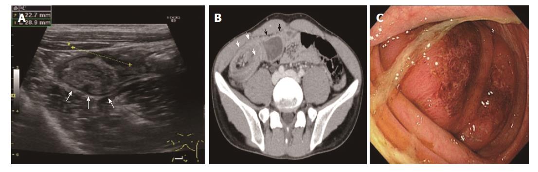
Figure 1 Target sign on abdominal ultrasonography (A) and contrast-enhanced computed tomography scan (B).
The so-called bowel-within-bowel configuration, in which the layers of the bowel are duplicated, thereby forming concentric rings, is seen (white arrows). Dilatation of the terminal ileum is observed (black arrows). C: A colonoscopic view of the intussusception. Edematous colonic mucosa was identified on the lead point of intussusception. No neoplastic lesion was detected, even after reduction of intussusception.
- Citation: Yamamoto T, Tajima Y, Hyakudomi R, Hirayama T, Taniura T, Ishitobi K, Hirahara N. Case of colonic intussusception secondary to mobile cecum syndrome repaired by laparoscopic cecopexy using a barbed wound suture device. World J Gastroenterol 2017; 23(35): 6534-6539
- URL: https://www.wjgnet.com/1007-9327/full/v23/i35/6534.htm
- DOI: https://dx.doi.org/10.3748/wjg.v23.i35.6534