Copyright
©The Author(s) 2017.
World J Gastroenterol. Sep 14, 2017; 23(34): 6365-6370
Published online Sep 14, 2017. doi: 10.3748/wjg.v23.i34.6365
Published online Sep 14, 2017. doi: 10.3748/wjg.v23.i34.6365
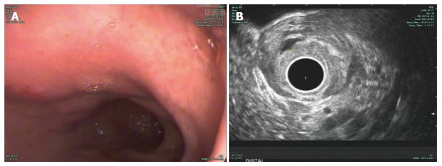
Figure 3 Esophagogastroduodenoscopy and endoscopic ultrasonography surveillance 5 mo later.
A: EGD surveillance 5 mo after stent insertion; B: Radial EUS confirmed the presence of a small anechoic cystic lesion measuring 3 mm × 5 mm. EUS: Endoscopic ultrasonography; EGD: Esophagogastroduodenoscopy.
- Citation: Jin HB, Lu L, Yang JF, Lou QF, Yang J, Shen HZ, Tang XW, Zhang XF. Interventional endoscopic ultrasound for a symptomatic pseudocyst secondary to gastric heterotopic pancreas. World J Gastroenterol 2017; 23(34): 6365-6370
- URL: https://www.wjgnet.com/1007-9327/full/v23/i34/6365.htm
- DOI: https://dx.doi.org/10.3748/wjg.v23.i34.6365


