Copyright
©The Author(s) 2017.
World J Gastroenterol. Sep 14, 2017; 23(34): 6365-6370
Published online Sep 14, 2017. doi: 10.3748/wjg.v23.i34.6365
Published online Sep 14, 2017. doi: 10.3748/wjg.v23.i34.6365
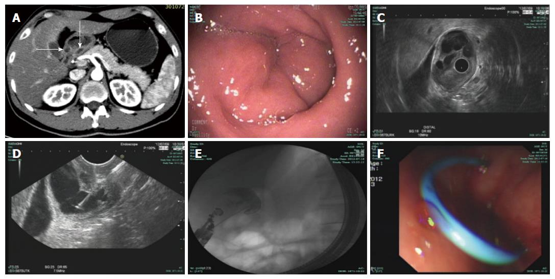
Figure 1 Computed tomography scan and endoscopic treatment of the lesion.
A: Contrast-enhanced CT revealed manifestations of a normal pancreas and an ill-defined enhancing mass (arrows) at the gastric antrum that caused circumferential narrowing of the pyloric channel; B: EGD showed a narrowed pyloric channel with normal mucosa; C: Radial EUS imaging confirmed the presence of a cystic lesion measuring 21 mm × 25 mm that originated from the third layer with anechoic contents and debris; D: EUS-guided fine needle aspiration; E: Cyst cavity radiography and guide-wire exchange; F: Successful insertion of a single pigtail stent (5 Fr × 4 cm). CT: Computed tomography; EUS: Endoscopic ultrasonography; EGD: Esophagogastroduodenoscopy.
- Citation: Jin HB, Lu L, Yang JF, Lou QF, Yang J, Shen HZ, Tang XW, Zhang XF. Interventional endoscopic ultrasound for a symptomatic pseudocyst secondary to gastric heterotopic pancreas. World J Gastroenterol 2017; 23(34): 6365-6370
- URL: https://www.wjgnet.com/1007-9327/full/v23/i34/6365.htm
- DOI: https://dx.doi.org/10.3748/wjg.v23.i34.6365


