Copyright
©The Author(s) 2017.
World J Gastroenterol. Sep 14, 2017; 23(34): 6350-6356
Published online Sep 14, 2017. doi: 10.3748/wjg.v23.i34.6350
Published online Sep 14, 2017. doi: 10.3748/wjg.v23.i34.6350
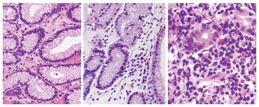
Figure 3 Biopsy for gastritis.
Gastric tissue biopsies were taken to compare the degree of gastritis: A: grade 0 [hematoxylin and eosin (HE) staining, × 200], normal mucosa with small amount of lymphocytes and transparent microscopic field; B: grade 1 (HE, × 200), intermediate between grades 0 and 2 with a moderate amount of lymphocytes or other kinds of inflammatory cells; C: grade 2 (HE, × 400), acute inflammation with fully infiltrated tissue by lymphocytes or other kinds of inflammatory cells.
- Citation: Yang D, He L, Tong WH, Jia ZF, Su TR, Wang Q. Randomized controlled trial of uncut Roux-en-Y vs Billroth II reconstruction after distal gastrectomy for gastric cancer: Which technique is better for avoiding biliary reflux and gastritis? World J Gastroenterol 2017; 23(34): 6350-6356
- URL: https://www.wjgnet.com/1007-9327/full/v23/i34/6350.htm
- DOI: https://dx.doi.org/10.3748/wjg.v23.i34.6350


