Copyright
©The Author(s) 2017.
World J Gastroenterol. Aug 21, 2017; 23(31): 5817-5822
Published online Aug 21, 2017. doi: 10.3748/wjg.v23.i31.5817
Published online Aug 21, 2017. doi: 10.3748/wjg.v23.i31.5817
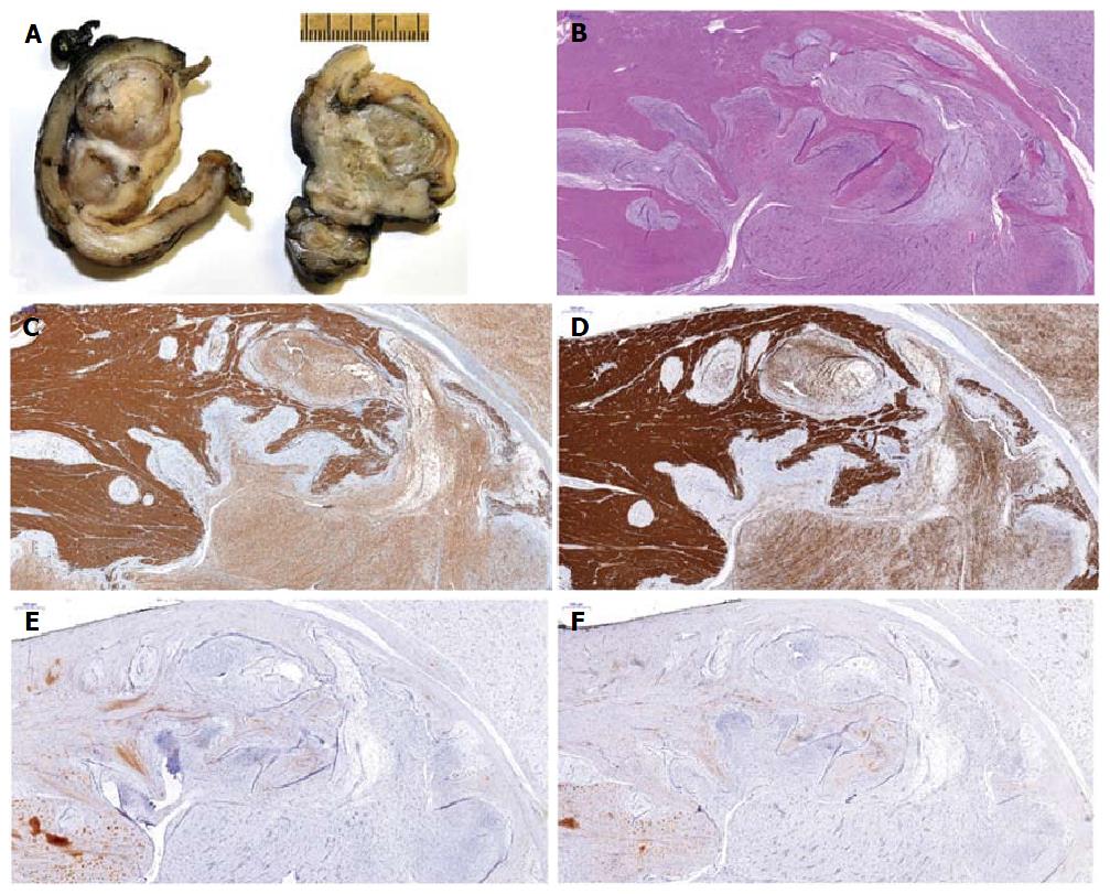
Figure 3 Macroscopic, microscopic and immunohistochemical features of plexiform fibromyxoma.
A: Cut section showing multinodular, solid glistening translucid tumor within the gastric wall and subserosa; B: Histological analysis of the tumor showing composition of ovoid to spindled-shaped cells in a myxoid or fibromyxoid stroma with a plexiform intramural growth pattern; C-F: Immunohistochemical analysis showing positivity for alpha-smooth muscle actin (C) and h-caldesmon (D), but negativity for KIT (E) and DOG1 (F).
- Citation: Szurian K, Till H, Amerstorfer E, Hinteregger N, Mischinger HJ, Liegl-Atzwanger B, Brcic I. Rarity among benign gastric tumors: Plexiform fibromyxoma - Report of two cases. World J Gastroenterol 2017; 23(31): 5817-5822
- URL: https://www.wjgnet.com/1007-9327/full/v23/i31/5817.htm
- DOI: https://dx.doi.org/10.3748/wjg.v23.i31.5817


