Copyright
©The Author(s) 2017.
World J Gastroenterol. Aug 21, 2017; 23(31): 5798-5808
Published online Aug 21, 2017. doi: 10.3748/wjg.v23.i31.5798
Published online Aug 21, 2017. doi: 10.3748/wjg.v23.i31.5798
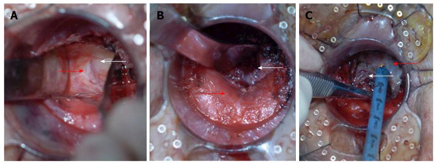
Figure 2 Dissection of the distal mesorectum in retrograde transanal total mesorectal excision.
A: Bilateral mobilization of the distal rectum. Dissection is started along the natural boundary between the surface of the levator ani muscle and mesorectum toward the pelvic cavity assisted. The two retractors are inserted into this space and used to expand it. The distance of bilateral mobilization toward the pelvic cavity could reach 10 cm according to the length of the retractors; B: Posterior mobilization of the distal rectum. The hiatal ligament is cut off after sharp dissection along the natural boundary between the surface of the levator ani muscle and mesorectum with an electrocautery or ultracision harmonic scalpel; C: Anterior mobilization of the distal rectum. The rectourethral muscle is cut off, and the Denonvilliers fascia is sharply dissected between the anterior and posterior lobes. White arrows indicate the rectum and its mesorectum, and red arrow indicates the levator ani muscle or Denonvilliers fascia.
- Citation: Xu C, Song HY, Han SL, Ni SC, Zhang HX, Xing CG. Simple instruments facilitating achievement of transanal total mesorectal excision in male patients. World J Gastroenterol 2017; 23(31): 5798-5808
- URL: https://www.wjgnet.com/1007-9327/full/v23/i31/5798.htm
- DOI: https://dx.doi.org/10.3748/wjg.v23.i31.5798


