Copyright
©The Author(s) 2017.
World J Gastroenterol. Aug 14, 2017; 23(30): 5619-5633
Published online Aug 14, 2017. doi: 10.3748/wjg.v23.i30.5619
Published online Aug 14, 2017. doi: 10.3748/wjg.v23.i30.5619
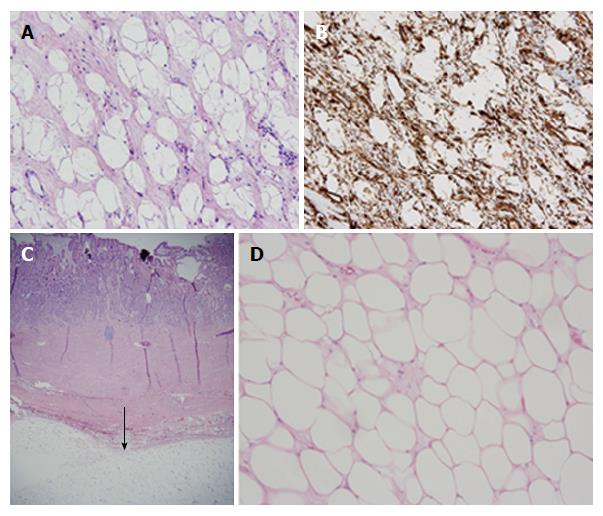
Figure 4 Histopathologic findings in gastrectomy specimens in two patients with giant gastric lipomas.
A: Patient 1-standard histochemistry. Medium power photomicrograph of a hematoxylin and eosin stain of a tissue section from the resected gastric mass in patient 1 reveals large adipocytes filled with clear, homogeneous, cytoplasm and tiny, compressed, peripheral nuclei. No lipoblasts are detected. Note the spindle-shaped stroma surrounding the adipocytes, findings consistent with spindle cell lipoma, as proven by immunohistochemistry (B); B: Patient 1-immunohistochemistry. Medium power photomicrograph of immunohistochemistry, using an antibody to CD34, reveals within tumor in patient 1 extensive staining in a spindly pattern of the stroma surrounding characteristically clear adipocytes, a characteristic staining pattern for spindle-shaped lipoma; C: Patient 2-standard histochemistry-low power. Low power photomicrograph of a hematoxylin and eosin stain of a tissue section from resected gastric mass in patient 2 reveals a well-circumscribed, submucosal layer composed of adipocytes with clear cytoplasm (arrow) and scant loose, myxoid stroma; D: Patient 2-standard histochemistry-medium power. Medium power photomicrograph of a hematoxylin and eosin stain of a tissue section from resected gastric mass in patient 2 reveals sheets of large, adipocytes filled with clear, homogeneous, cytoplasm and tiny, compressed, peripheral nuclei, with scant loose, myxoid stroma. No lipoblasts are detected. These histologic findings are characteristic of lipomas.
- Citation: Cappell MS, Stevens CE, Amin M. Systematic review of giant gastric lipomas reported since 1980 and report of two new cases in a review of 117110 esophagogastroduodenoscopies. World J Gastroenterol 2017; 23(30): 5619-5633
- URL: https://www.wjgnet.com/1007-9327/full/v23/i30/5619.htm
- DOI: https://dx.doi.org/10.3748/wjg.v23.i30.5619


