Copyright
©The Author(s) 2017.
World J Gastroenterol. Aug 14, 2017; 23(30): 5619-5633
Published online Aug 14, 2017. doi: 10.3748/wjg.v23.i30.5619
Published online Aug 14, 2017. doi: 10.3748/wjg.v23.i30.5619
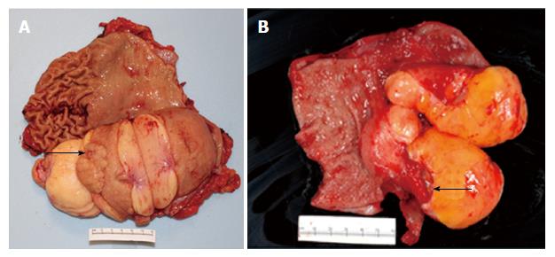
Figure 3 Gross pathologic findings in gastrectomy specimens in two patients with giant gastric lipomas.
A: Patient 1. Patient 1 presented with acute melena and hemoglobin decline and had an ulcer detected at esophagogastroduodenoscopy (EGD) within a huge, lipomatous gastric mass. Gross pathologic view of the gastrectomy specimen after it is opened to expose the luminal surface shows a well-circumscribed, lobulated, 14.5 cm × 9.0 cm × 7.5 cm lipomatous mass extending from the gastric body (left) to antrum (right). A small ulcer (round depression, arrow) is present on the mucosa overlying the lipomatous mass. Normal gastric rugae are present above the mass on the upper left, but have been effaced on the upper right, likely because of chronic compression/pressure from the giant lipomatous mass located below (on the contralateral gastric wall before opening the stomach). Vertical incisions show a homogeneous yellow-tan cut surface, indicative of a lipomatous tumor; B: Patient 2. Patient 2 presented with melena for 3 d, orthostatic dizziness, and a hemoglobin decline to 7.1 g/dL requiring transfusion of 2 units of packed erythrocytes and had at EGD a large, yellowish, smooth, well-circumscribed antral mass. Gross pathologic view of distal gastrectomy specimen after it is opened to expose the luminal surface shows normal gastric antral tissue at left and a lobulated, well-circumscribed, yellow-tan, 9.0 cm × 6.0 cm × 3.5 cm lipomatous tumor at right, with a deep, clean-based, ulcer (arrow) on the mucosa overlying the mass.
- Citation: Cappell MS, Stevens CE, Amin M. Systematic review of giant gastric lipomas reported since 1980 and report of two new cases in a review of 117110 esophagogastroduodenoscopies. World J Gastroenterol 2017; 23(30): 5619-5633
- URL: https://www.wjgnet.com/1007-9327/full/v23/i30/5619.htm
- DOI: https://dx.doi.org/10.3748/wjg.v23.i30.5619


