Copyright
©The Author(s) 2017.
World J Gastroenterol. Aug 14, 2017; 23(30): 5619-5633
Published online Aug 14, 2017. doi: 10.3748/wjg.v23.i30.5619
Published online Aug 14, 2017. doi: 10.3748/wjg.v23.i30.5619
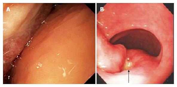
Figure 1 Findings at esophagogastroduodenoscopy in two patients with giant gastric lipomas.
A: Patient 1. Esophagogastroduodenoscopy (EGD) in a 63-year-old male who presented with melena and a hemoglobin decline to 6.2 g/dL that required transfusion of 2 units of packed erythrocytes, showing the distal body and antrum with a huge mass folded upon itself occupying most of the lumen and an 8 mm wide, nonbleeding, acute mucosal ulcer without stigmata of recent hemorrhage embedded deep in the valley (fold) between the right and left parts of the mass. The ulcer was attributed to friction from the opposing surface. The mass was 13-cm-wide, submucosal, yellowish, and covered by smooth mucosa except for focal ulceration, findings consistent with a gastric lipoma; B: Patient 2. EGD in a 78-year-old-woman, who presented with melena for 3 d, orthostatic dizziness, and a hemoglobin decline to 7.1 g/dL requiring transfusion of 2 units of packed erythrocytes, revealed an acute 1-cm-wide prepyloric ulcer (arrow) with a white exudate but without stigmata of recent hemorrhage between the right and left lobes of a large, well-demarcated, submucosal, mass covered by otherwise normal, superficial mucosa. This endoscopic photograph shows only a part of the mass.
- Citation: Cappell MS, Stevens CE, Amin M. Systematic review of giant gastric lipomas reported since 1980 and report of two new cases in a review of 117110 esophagogastroduodenoscopies. World J Gastroenterol 2017; 23(30): 5619-5633
- URL: https://www.wjgnet.com/1007-9327/full/v23/i30/5619.htm
- DOI: https://dx.doi.org/10.3748/wjg.v23.i30.5619


