Copyright
©The Author(s) 2017.
World J Gastroenterol. Aug 7, 2017; 23(29): 5304-5312
Published online Aug 7, 2017. doi: 10.3748/wjg.v23.i29.5304
Published online Aug 7, 2017. doi: 10.3748/wjg.v23.i29.5304
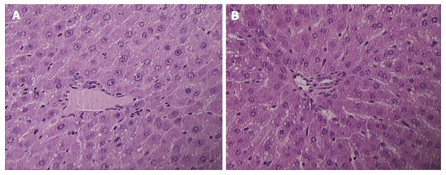
Figure 2 Presentation of celecoxib-induced liver lesions at 48 h.
Controls presented with pronounced microvesicullar and macrovesicullar steatosis, dilated sinusoids, and piecemeal necrosis (A); BPC 157 rats presenting with minimal microvesicullar steatosis and no necrosis (B). HE × 40.
- Citation: Drmic D, Kolenc D, Ilic S, Bauk L, Sever M, Zenko Sever A, Luetic K, Suran J, Seiwerth S, Sikiric P. Celecoxib-induced gastrointestinal, liver and brain lesions in rats, counteraction by BPC 157 or L-arginine, aggravation by L-NAME. World J Gastroenterol 2017; 23(29): 5304-5312
- URL: https://www.wjgnet.com/1007-9327/full/v23/i29/5304.htm
- DOI: https://dx.doi.org/10.3748/wjg.v23.i29.5304


