Copyright
©The Author(s) 2017.
World J Gastroenterol. Jul 28, 2017; 23(28): 5115-5126
Published online Jul 28, 2017. doi: 10.3748/wjg.v23.i28.5115
Published online Jul 28, 2017. doi: 10.3748/wjg.v23.i28.5115
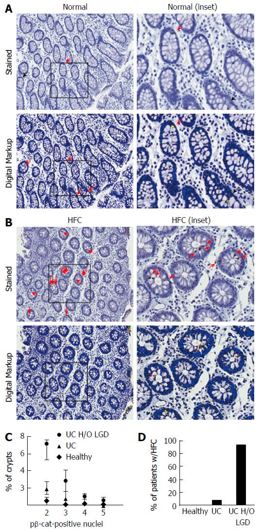
Figure 5 Crypts with elevated pβ-cat staining identify ulcerative colitis patients at a high risk for low grade dysplasia.
A: Representative images of pβ-cat staining and the digital markup in colon biopsies of UC patients with normal pβ-cat distribution (benign disease); and B: HFC. Red arrows indicated pβ-cat-positive nuclei identified by eye; black arrows indicate false positive or negative nuclei properly identified by digital analysis; C: Statistical distribution of pβ-cat-positive nuclei in normal biopsies showing that crypts with 2 more positive pβ-cat-positive nuclei are rarely detected in healthy patients (diamonds, n = 3) or patients with benign, active UC (triangles, n = 12) but are more frequent in UC patients with a previous history of LGD (circles, n = 16). Data are shown as the percent of crypt cross-sections containing pβ-cat-positive nuclei; D: Biopsies from healthy controls, benign UC, and UC patients with a history of LGD were analyzed for the presence of HFC. UC: Ulcerative colitis; LGD: Low grade dysplasia; HFC: High frequency crypts.
- Citation: Bradford EM, Thompson CA, Goretsky T, Yang GY, Rodriguez LM, Li L, Barrett TA. Myo-inositol reduces β-catenin activation in colitis. World J Gastroenterol 2017; 23(28): 5115-5126
- URL: https://www.wjgnet.com/1007-9327/full/v23/i28/5115.htm
- DOI: https://dx.doi.org/10.3748/wjg.v23.i28.5115


