Copyright
©The Author(s) 2017.
World J Gastroenterol. Jul 7, 2017; 23(25): 4615-4623
Published online Jul 7, 2017. doi: 10.3748/wjg.v23.i25.4615
Published online Jul 7, 2017. doi: 10.3748/wjg.v23.i25.4615
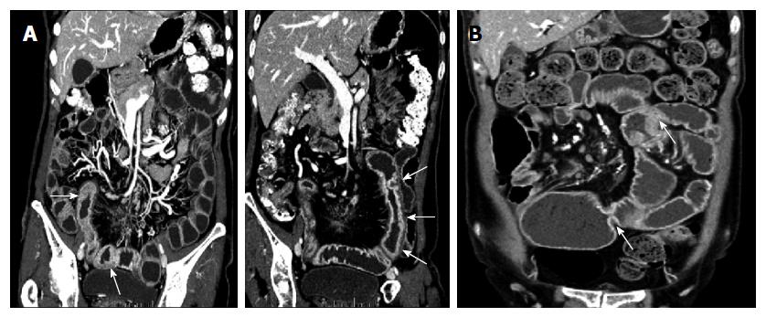
Figure 3 A 49-year-old woman with abdominal pain.
A: Coronal reformatted images of computed tomography (CT) enterography show long segmental bowel wall thickening with layered enhancement pattern, involving distal jejunum and proximal ileum. B: This bowel wall thickening of the small intestine is not noted on the coronal reformatted image of CT enterography four years later, and instead, several short-segmental strictures (arrows) have developed in the same bowel loop.
- Citation: Hwang J, Kim JS, Kim AY, Lim JS, Kim SH, Kim MJ, Kim MS, Song KD, Woo JY. Cryptogenic multifocal ulcerous stenosing enteritis: Radiologic features and clinical behavior. World J Gastroenterol 2017; 23(25): 4615-4623
- URL: https://www.wjgnet.com/1007-9327/full/v23/i25/4615.htm
- DOI: https://dx.doi.org/10.3748/wjg.v23.i25.4615


