Copyright
©The Author(s) 2017.
World J Gastroenterol. Jun 28, 2017; 23(24): 4369-4380
Published online Jun 28, 2017. doi: 10.3748/wjg.v23.i24.4369
Published online Jun 28, 2017. doi: 10.3748/wjg.v23.i24.4369
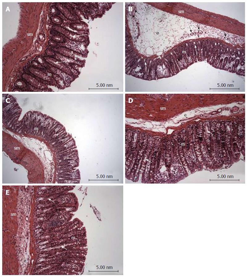
Figure 2 Photomicrography (hematoxylin and eosin) of colon samples from different experimental groups.
Mucosa (m), tubular glands (white arrows), blood vessels (black arrow), submucosa (sm) and edema (e). In A (non-colitic group): the mucosa (m) contains numerous straight tubular glands (white arrows) with many lightly stained goblet cells; the crypt (black arrow), submucosa (sm) and luminal epithelium was intact with a typical morphology; In B (TNBS control group): tubular glands (white arrows) were reduced and the goblet cells were atypical; a disruption with extensive edema (e) and blood vessels (black arrow) can be observed. In PACO2-treated groups, colon cytoarchitecture was recovering and included the restoration of tubular glands (white arrows) containing goblet cells, especially at the doses of 50 mg/kg (D) and 100 mg/kg (E); edema (e) was significantly reduced in animals treated with at doses of 50 mg/kg (D) and 100 mg/kg (E) and in a lower proportion in animals treated with doses of 25 mg/kg (C), when compared to the TNBS control group (B).
- Citation: Almeida Junior LD, Quaglio AEV, de Almeida Costa CAR, Di Stasi LC. Intestinal anti-inflammatory activity of Ground Cherry (Physalis angulata L.) standardized CO2 phytopharmaceutical preparation. World J Gastroenterol 2017; 23(24): 4369-4380
- URL: https://www.wjgnet.com/1007-9327/full/v23/i24/4369.htm
- DOI: https://dx.doi.org/10.3748/wjg.v23.i24.4369


