Copyright
©The Author(s) 2017.
World J Gastroenterol. Jun 14, 2017; 23(22): 4064-4071
Published online Jun 14, 2017. doi: 10.3748/wjg.v23.i22.4064
Published online Jun 14, 2017. doi: 10.3748/wjg.v23.i22.4064
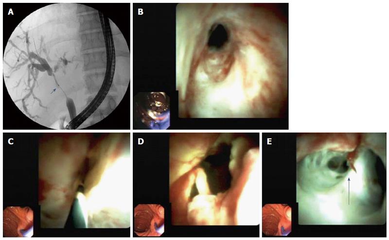
Figure 2 Non-anastomotic strictures in seven.
A: High-grade, long-segment stenosis of the main bile duct between biliary anastomosis and the hilus region. The biliary hilus appears to be involved in the stenosis; B: Cholangioscopic image of biliary anastomosis with slight stenosis; C: High-grade stricture of the bile duct beyond the biliary stricture that could not be passed using a cholangioscope; D: Bile duct above the biliary anastomosis after balloon dilation with marked erythema and polypoid growth of the bile duct wall, and signs of pronounced inflammation; E: Bile duct at the height of the hilus (arrow) with inflammation involving the left hepatic bile duct (right side of the image).
- Citation: Hüsing-Kabar A, Heinzow HS, Schmidt HHJ, Stenger C, Gerth HU, Pohlen M, Thölking G, Wilms C, Kabar I. Single-operator cholangioscopy for biliary complications in liver transplant recipients. World J Gastroenterol 2017; 23(22): 4064-4071
- URL: https://www.wjgnet.com/1007-9327/full/v23/i22/4064.htm
- DOI: https://dx.doi.org/10.3748/wjg.v23.i22.4064


