Copyright
©The Author(s) 2017.
World J Gastroenterol. Jun 7, 2017; 23(21): 3928-3933
Published online Jun 7, 2017. doi: 10.3748/wjg.v23.i21.3928
Published online Jun 7, 2017. doi: 10.3748/wjg.v23.i21.3928
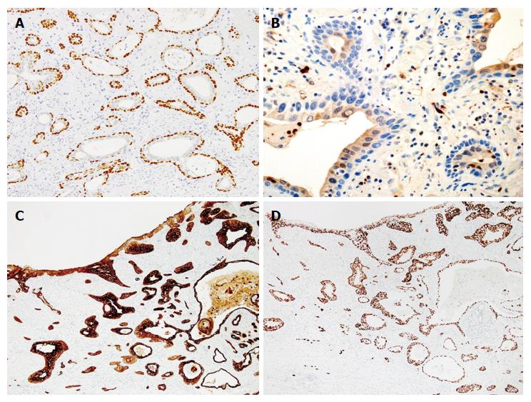
Figure 3 Immunohistochemical examinations.
A: p63 (magnification 200 ×); B: S-100 (magnification 400 ×); C: CK7 (magnification 100 ×); D: p53 (magnification 100 ×). The outer layer cells of the neoplastic tubules were reactive for p63 and CK7, but negative for S100. The inner layer cells were immunopositive for S100, CK7, but negative for p63. Strong expressions of p53 and CK7 were observed in the small area of the surface epithelium connecting with the invasive carcinoma component. Both the inner and outer layer cells in the invasive component also overexpressed p53 protein.
- Citation: Tamura H, Saiki H, Amano T, Yamamoto M, Hayashi S, Ando H, Doi R, Nishida T, Yamamoto K, Adachi S. Esophageal carcinoma originating in the surface epithelium with immunohistochemically proven esophageal gland duct differentiation: A case report. World J Gastroenterol 2017; 23(21): 3928-3933
- URL: https://www.wjgnet.com/1007-9327/full/v23/i21/3928.htm
- DOI: https://dx.doi.org/10.3748/wjg.v23.i21.3928


