Copyright
©The Author(s) 2017.
World J Gastroenterol. May 28, 2017; 23(20): 3752-3757
Published online May 28, 2017. doi: 10.3748/wjg.v23.i20.3752
Published online May 28, 2017. doi: 10.3748/wjg.v23.i20.3752
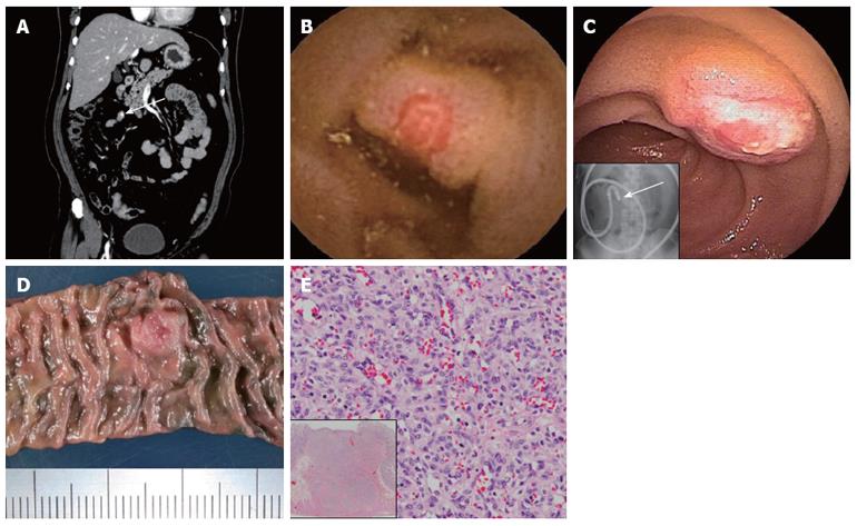
Figure 3 Evaluation of clinical finding (case 2).
Early-phase contrast-enhanced computed tomography reveals small nodule enhancement in the ileum (arrow) (A). Video capsule endoscopy (B) and double-balloon enteroscopy (C) show a submucosal tumor-like raised lesion with central erosion in the lower ileum. The lesion in the jejunum was disclosed 145 min after capsule ingestion (pylorus passage at 140 min) (B), inset indicates fluoroscopic localization of target at the end of the endoscope insertion (arrow) (C). Surgical specimen from the small intestine, including the indicated lesion with India ink tattooing (D). Histological finding (H-E stain) in the resected specimen show cirumferential capillary growth without atypia from the mucosa to the muscle. Inset shows a low-power filed view (E).
- Citation: Takase N, Fukui K, Tani T, Nishimura T, Tanaka T, Harada N, Ueno K, Takamatsu M, Nishizawa A, Okamura A, Kaneda K. Preoperative detection and localization of small bowel hemangioma: Two case reports. World J Gastroenterol 2017; 23(20): 3752-3757
- URL: https://www.wjgnet.com/1007-9327/full/v23/i20/3752.htm
- DOI: https://dx.doi.org/10.3748/wjg.v23.i20.3752


