Copyright
©The Author(s) 2017.
World J Gastroenterol. May 28, 2017; 23(20): 3752-3757
Published online May 28, 2017. doi: 10.3748/wjg.v23.i20.3752
Published online May 28, 2017. doi: 10.3748/wjg.v23.i20.3752
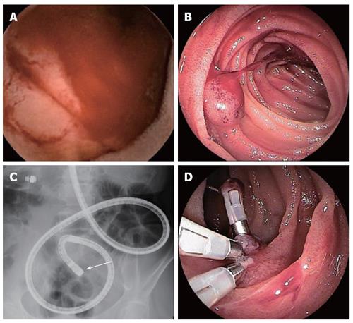
Figure 1 Evaluation of endoscopic findings (case 1).
Video capsule endoscopy (A) and double-enteroscopy (B and C) show a raised lesion with smooth surface in the upper jejunum, and double-balloon enterscopy showed spout bleeding of the lesion. The lesion in the jejunum was disclosed 29 min after capsule ingestion (pylonus passage at 16 min) (A). Detailed localization of the target lesion using fluoroscopy is shown by the end of endoscopic insertion (arrow) (C). The lesion underwent endoscopic hemostatic clipping (D).
- Citation: Takase N, Fukui K, Tani T, Nishimura T, Tanaka T, Harada N, Ueno K, Takamatsu M, Nishizawa A, Okamura A, Kaneda K. Preoperative detection and localization of small bowel hemangioma: Two case reports. World J Gastroenterol 2017; 23(20): 3752-3757
- URL: https://www.wjgnet.com/1007-9327/full/v23/i20/3752.htm
- DOI: https://dx.doi.org/10.3748/wjg.v23.i20.3752


