
















©The Author(s) 2017.
World J Gastroenterol. May 28, 2017; 23(20): 3664-3674
Published online May 28, 2017. doi: 10.3748/wjg.v23.i20.3664
Published online May 28, 2017. doi: 10.3748/wjg.v23.i20.3664
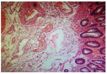
Figure 5 Subepithelial vessels of resected grades III and IV hemorrhoid tissues were manifested by obvious structural impairment and retrograde and ruptured changes of internal elastic lamina.
Arteriovenous fistulas and venous dilatation were obvious in the anal cushion of hemorhoidal tissues (white arrow, magnification × 80); A: Artery, V: Vein, L: Lymphatic duct.
- Citation: Aimaiti A, A Ba Bai Ke Re MMTJ, Ibrahim I, Chen H, Tuerdi M, Mayinuer. Sonographic appearance of anal cushions of hemorrhoids. World J Gastroenterol 2017; 23(20): 3664-3674
- URL: https://www.wjgnet.com/1007-9327/full/v23/i20/3664.htm
- DOI: https://dx.doi.org/10.3748/wjg.v23.i20.3664