Copyright
©The Author(s) 2017.
World J Gastroenterol. Jan 14, 2017; 23(2): 297-305
Published online Jan 14, 2017. doi: 10.3748/wjg.v23.i2.297
Published online Jan 14, 2017. doi: 10.3748/wjg.v23.i2.297
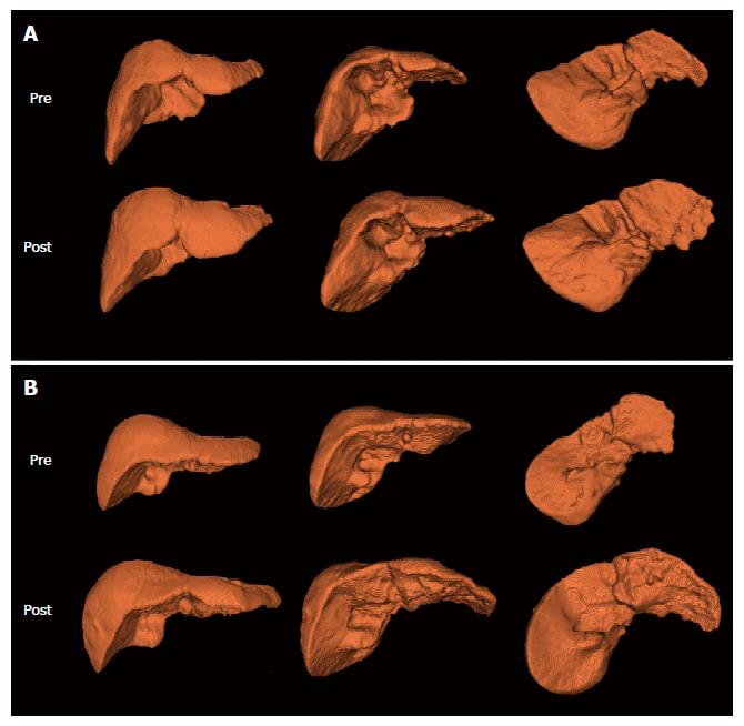
Figure 2 Representative 3-dimentional reconstruction images in patients with (A) compensated liver cirrhosis and (B) decompensated liver cirrhosis.
Among 6 images for each patient, upper images are baseline (pre-NUCs therapy) images and lower images are follow-up (post-NUCs therapy) images in different views (anterior, anteroinferomedial, and posteroinferomedial). NUCs: Nucleos(t)ide analogues.
- Citation: Lee CH, Kim IH, Moon JC, Seo SY, Kim SH, Kim SW, Lee SO, Lee ST, Kim DG, Yang JD, Yu HC. 3-Dimensional liver volume assessment in patients with hepatitis B virus-related liver cirrhosis during long-term oral nucleos(t)ide analogues therapy. World J Gastroenterol 2017; 23(2): 297-305
- URL: https://www.wjgnet.com/1007-9327/full/v23/i2/297.htm
- DOI: https://dx.doi.org/10.3748/wjg.v23.i2.297


