Copyright
©The Author(s) 2017.
World J Gastroenterol. May 21, 2017; 23(19): 3468-3479
Published online May 21, 2017. doi: 10.3748/wjg.v23.i19.3468
Published online May 21, 2017. doi: 10.3748/wjg.v23.i19.3468
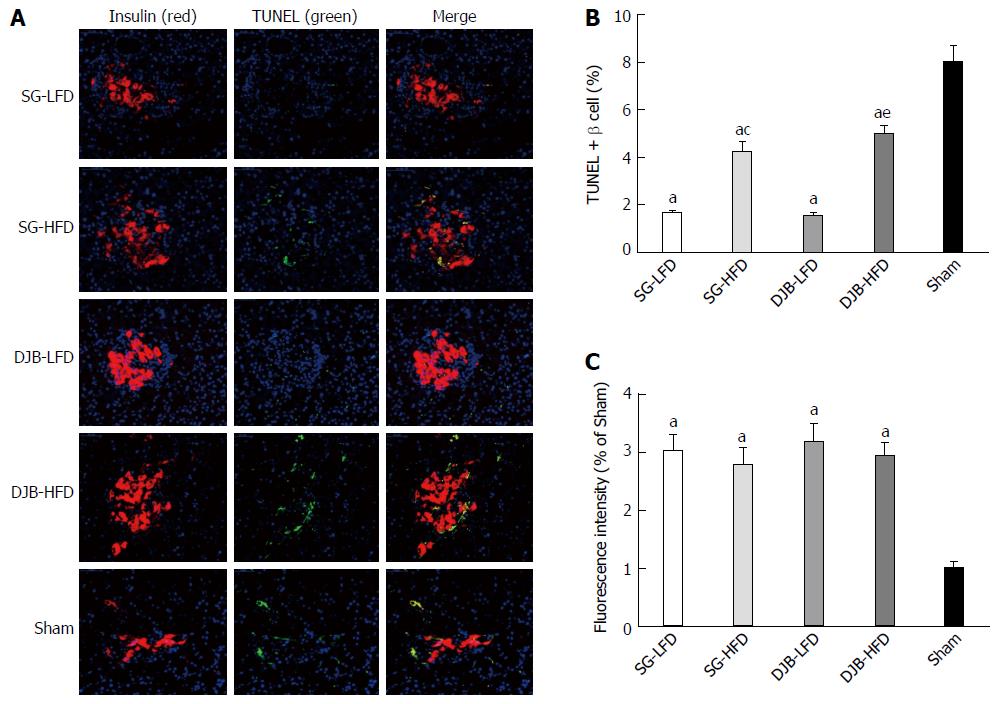
Figure 5 Pancreatic beta cell analysis.
A: TUNEL-positive beta cell shown by immunofluorescence; B: and the percentage of TUNEL-positive beta cells was calculated. The two surgery group had fewer TUNEL-positive beta cells than the Sham group, and HFD group made the percentage higher than in the LFD group; C: The fluorescence intensity did not differ significantly between the four surgery groups (P > 0.05), but was higher than in the Sham group. aP < 0.05 vs Sham group at 12 wk postoperatively; cP < 0.05 vs SG-LFD group at 12 wk postoperatively; eP < 0.05 vs DJB-LFD group at 12 wk postoperatively. TUNEL: Transferase-mediated dUTP-biotin 3’ nick end-labeling; SG: Sleeve gastrectomy; SG-LFD: SG rats with common low-fat diet; SG-HFD: SG with high-fat diet; DJB: Duodenal-jejunal bypass; DJB-LFD: DJB rats with LFD; DJB-HFD: DJB rats with HFD.
- Citation: Liu T, Zhong MW, Liu Y, Sun D, Wei M, Huang X, Cheng YG, Wu QZ, Wu D, Zhang XQ, Wang KX, Hu SY, Liu SZ. Diabetes recurrence after metabolic surgeries correlates with re-impaired insulin sensitivity rather than beta-cell function. World J Gastroenterol 2017; 23(19): 3468-3479
- URL: https://www.wjgnet.com/1007-9327/full/v23/i19/3468.htm
- DOI: https://dx.doi.org/10.3748/wjg.v23.i19.3468


